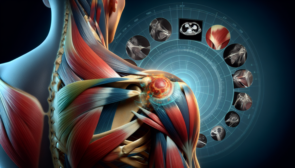
Rotator cuff tears can be a painful and debilitating condition affecting the shoulder joint. This common injury occurs when one or more of the tendons connecting the shoulder muscles to the upper arm bone become damaged or torn. Understanding how to diagnose and treat a rotator cuff tear is crucial for healthcare professionals and patients alike, as proper management can make a significant difference in recovery and long-term shoulder function.
This article delves into the classification of rotator cuff tears, explores the challenges in diagnosis, and outlines effective solutions. It also discusses the importance of personalized treatment planning, highlighting various options available to patients. Additionally, the article covers essential aspects of recovery and rehabilitation, providing valuable insights into the journey from injury to restored shoulder health. By examining these key areas, readers will gain a comprehensive understanding of rotator cuff tear management.
Rotator Cuff Tear Classification
Rotator cuff tears can be classified based on their size, location, and extent of the tear. The most common categories include partial and full-thickness tears, as well as acute and chronic tears.
Partial vs. Full-Thickness Tears
Partial tears, also known as incomplete tears, occur when the tendon is damaged but not completely severed. The tendon remains partially attached to the bone, although it may be thinned or frayed. In contrast, full-thickness tears, or complete tears, involve the complete detachment of the tendon from the bone. Full-thickness tears can be further classified as full-thickness incomplete tears, where only a small part of the tendon is detached, or full-thickness complete tears, where there is essentially a hole in the tendon.
RELATED: Albinism: In-Depth Look at Causes, Symptoms, and Types
Acute vs. Chronic Tears
Rotator cuff tears can also be categorized as acute or chronic. Acute tears typically result from a sudden, traumatic event, such as a fall or lifting a heavy object with a jerking motion. These tears can occur in conjunction with other injuries, like a broken collarbone or dislocated shoulder. Chronic tears, on the other hand, develop gradually over time due to wear and tear on the tendon. They are more common in individuals over the age of 40 and those who engage in repetitive overhead activities.
Size and Location Considerations
The size of a rotator cuff tear is an important factor in determining the severity of the injury and the most appropriate treatment approach. Tears are often measured in centimeters, with larger tears generally requiring more extensive treatment. The location of the tear within the rotator cuff tendons also plays a role in the classification and management of the injury. Tears can occur in the supraspinatus, infraspinatus, teres minor, or subscapularis tendons, with each location presenting unique challenges for repair and rehabilitation.
Diagnostic Challenges and Solutions
Diagnosing rotator cuff tears can be challenging due to the complex anatomy of the shoulder and the presence of other conditions that mimic rotator cuff pathology. A thorough clinical evaluation is essential for an accurate diagnosis. During the physical exam, the physician should assess for pain with overhead activity, weakness on empty can and external rotation tests, and a positive impingement sign, as these findings have a high probability of indicating a rotator cuff tear.
Imaging modalities play a crucial role in confirming the diagnosis and determining the extent of the tear. Plain radiographs may show indirect signs of a massive rotator cuff tear, such as superior migration of the humeral head and narrowing of the acromiohumeral distance. MRI and ultrasonography are the preferred imaging tests for rotator cuff disorders, with both demonstrating high sensitivity and specificity for detecting full-thickness and partial-thickness tears. MRI provides a global assessment of all shoulder structures, while ultrasonography offers dynamic evaluation and higher spatial resolution.
Several differential diagnoses should be considered when evaluating a patient with suspected rotator cuff pathology. These include labral tears, glenohumeral instability, adhesive capsulitis, and referred pain from the cervical spine. Labral tears can present with shoulder pain, instability, and weakness, similar to rotator cuff tears. Glenohumeral instability is more common in younger patients and is often associated with a history of dislocation or subluxation events. Adhesive capsulitis is characterized by gradual onset of pain and global restriction of both active and passive range of motion. Referred pain from the cervical spine can mimic shoulder pathology, with pain and tenderness in the suprascapular area and reproduction of symptoms with cervical spine movement.
Personalized Treatment Planning
When developing a personalized treatment plan for a rotator cuff tear, several factors must be considered to optimize healing potential and functional outcomes. The patient’s age, tear size and chronicity, muscle quality, and tendon retraction are important variables that influence the decision between surgical repair and conservative management.
Older patients with chronic, massive tears and poor muscle quality may benefit more from an initial trial of physical therapy, as healing rates after surgical repair are significantly lower in this population. In contrast, younger patients with acute, traumatic tears or chronic tears larger than 1-1.5 cm without significant muscle atrophy or fatty infiltration should be considered for early surgical intervention to prevent irreversible changes and improve the likelihood of successful healing.
RELATED: What Are Adenomas? Key Facts and Information
For partial-thickness tears, small full-thickness tears, and patients with significant medical comorbidities, prolonged conservative treatment is often the preferred approach due to the limited risk of tear progression and good potential for functional improvement with physical therapy. Modifiable risk factors such as smoking cessation and optimizing blood glucose and cholesterol levels should be addressed to enhance healing potential.
If surgery is indicated, repair construct and rehabilitation protocol also play a role in treatment planning. Double-row repairs have demonstrated higher healing rates compared to single-row techniques, particularly for larger tears. Slower, less aggressive rehabilitation programs may reduce the risk of re-tear without compromising long-term range of motion and should be considered for most patients after rotator cuff repair.
Developing a personalized treatment plan requires careful consideration of patient-specific factors, tear characteristics, and surgeon preferences to determine the most appropriate approach for optimizing rotator cuff healing and functional outcomes in each individual case.
Recovery and Rehabilitation Essentials
Immediate Post-Treatment Care
Following rotator cuff repair surgery, patients must wear a sling for 4-6 weeks to immobilize the shoulder and protect the healing tissues. During this period, no active shoulder movements are allowed. Patients may remove the sling for showering and dressing but should avoid using the affected arm. Pain management is crucial in the early stages of recovery, and medications such as opioids, acetaminophen, and NSAIDs may be prescribed. Applying ice to the shoulder can also help reduce pain and inflammation.
Progressive Rehabilitation Phases
Rehabilitation after rotator cuff surgery progresses through several phases. In the first phase, passive range of motion exercises are introduced to prevent stiffness and maintain joint mobility. These exercises are performed by a physical therapist or an assisted device, with the patient’s muscles remaining relaxed. As healing progresses, active-assisted and active range of motion exercises are gradually incorporated. Strengthening exercises for the rotator cuff and surrounding muscles typically begin 6-12 weeks after surgery, depending on the extent of the tear and the quality of the repair. Resistance is progressively increased as strength improves.
RELATED: Antiphospholipid Syndrome: From Diagnosis to Treatment
Return to Activity Guidelines
The timeline for returning to daily activities and sports varies depending on the individual and the nature of the rotator cuff tear. Most patients can resume light activities and desk work within 2-4 weeks after surgery. Driving may be allowed once the sling is discontinued and the patient has adequate strength and range of motion. A return to sports typically occurs 4-6 months after surgery for non-contact activities and 6-12 months for contact or overhead sports. Adherence to the rehabilitation protocol and guidance from the surgeon and physical therapist are essential for a successful return to activity after rotator cuff repair.
Conclusion
Rotator cuff tears have a significant impact on shoulder function and quality of life. This article has explored the classification, diagnosis, treatment planning, and rehabilitation of these injuries. Understanding the different types of tears, from partial to full-thickness and acute to chronic, is crucial to develop an effective treatment strategy. The challenges in diagnosis highlight the need for a comprehensive approach, combining clinical evaluation with advanced imaging techniques to ensure accurate assessment.
Personalized treatment planning plays a key role in managing rotator cuff tears. By considering factors such as the patient’s age, tear characteristics, and overall health, healthcare providers can tailor interventions to maximize healing potential and functional outcomes. Whether opting for conservative management or surgical repair, the recovery process demands patience and dedication. A well-structured rehabilitation program, progressing through various phases and adhering to activity guidelines, is essential to restore shoulder function and prevent re-injury. This holistic approach to rotator cuff tear management aims to help patients regain strength, mobility, and confidence in their shoulder function.

