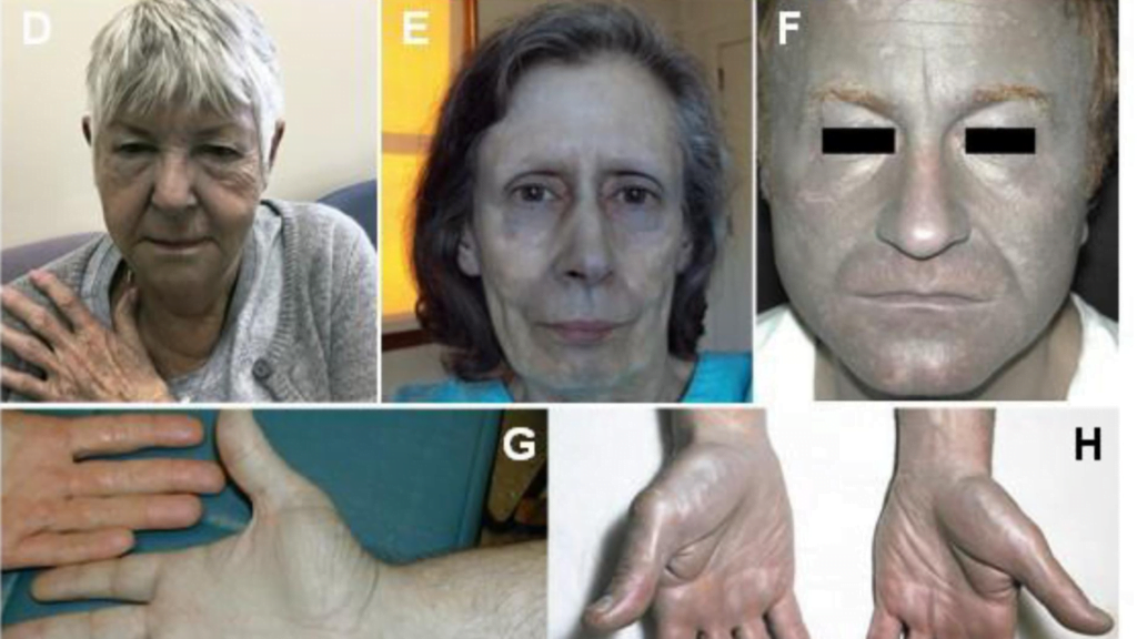
Argyria, a rare condition characterized by the skin acquiring a blue-gray hue, has piqued medical curiosity for centuries. Its distinctive discoloration, often resulting from prolonged exposure to silver compounds, raises questions about the balance between necessary treatment and potential cosmetic concerns. Understanding argyria, including its causes, symptoms, and treatments, is crucial for both medical professionals and those affected by the disease. This condition, while not widely known, epitomizes the intricate relationships between metals and human health, underscoring the importance of careful therapeutic use of silver-based products.
The subsequent sections of this article will delve into the chemistry and history of silver, elucidating how its medicinal applications can sometimes lead to argyria. The causes of argyria, pivotal for recognizing risk factors, will be discussed alongside its symptoms and diagnosis, providing a comprehensive overview of how this condition manifests and how it can be identified. The exploration will extend to the subtypes of argyria, its pathophysiology, and, importantly, the preventative measures and treatment options available. Through this detailed examination, the article aims to demystify argyria, offering insight into whether argyria is deadly, and how those affected can manage their symptoms and improve their quality of life.
Chemistry and History of Silver
Silver has held a significant role in various societies, not only as a material for crafting jewelry and minting coins but also for its applications in health and medicine. Historically, silver was used in its elemental form and as compounds in medical treatments, leveraging its antimicrobial properties. This section explores the dual roles of silver, both in economic and medicinal contexts, and how these have contributed to instances of argyria.
Economic and Cultural Significance of Silver
Silver’s versatility extends beyond its lustrous appeal in jewelry and artifacts. Economically, it has been a staple in the form of coins across various cultures, signifying wealth and stability. The expression “born with a silver spoon in his mouth” originally highlighted silver’s health benefits rather than wealth, as silver utensils were believed to prevent diseases in children. This belief underlines the historical recognition of silver’s antibacterial properties, which were appreciated long before the modern understanding of microbial pathogens.
Medicinal Applications of Silver
The oligodynamic effect of silver, a term describing the antimicrobial effects exerted by small amounts of heavy metals, underscores its continued relevance in medicine. Silver ions, released from silver-containing objects or solutions, are known to interact with bacterial membranes, causing irreversible damage. This antimicrobial property was historically exploited in various forms, including colloidal silver—a suspension of microscopic silver particles used as an internal medication.
Historical Use and the Onset of Argyria
By the mid-19th century, the medical community recognized that prolonged exposure to silver, whether through ingestion or inhalation, could lead to argyria, a condition marked by the skin turning grey or blue-grey. Workers in silver manufacturing facilities were particularly at risk, as were those using silver-containing products over extended periods. The decline in the use of colloidal silver in favor of antibiotics like penicillin in the 1940s marked a significant shift in medical treatments, although cases of argyria persisted due to exposure from other sources.
Silver in Modern Medical Applications and Argyria Cases
The use of silver in medical applications continued into the modern era, with documented cases of argyria resulting from silver used in medical instruments and as a food supplement in the form of colloidal silver. The condition primarily manifests in sun-exposed areas of the skin, where the deposited silver particles react to sunlight, enhancing the pigmentation. This reaction is similar to photographic development, where silver salts reduce to elemental silver under light exposure.
In summary, silver’s role in society and medicine has been multifaceted, contributing both to economic development and medical advancements. However, its use has also led to health complications such as argyria, highlighting the need for caution in its application. Understanding the chemistry and historical context of silver provides a foundation for comprehending the complexities of its benefits and risks.
Causes of Argyria
Prolonged Exposure to Silver
Argyria can develop from prolonged exposure to silver, particularly in the form of microscopic silver compounds that are absorbed into the body over time. This exposure is often seen in individuals who ingest silver particles through medications or dietary supplements that contain silver salts, colloidal silver, or silver acetate. The body’s natural ability to process small amounts of silver is overwhelmed when silver is ingested regularly, leading to the accumulation of silver in tissues, which then manifests as a blue to gray discoloration of the skin and mucous membranes.
High-Dose Exposure to Silver
While prolonged exposure is a common cause, argyria can also occur from a single, high-dose exposure to silver compounds. This type of exposure can lead to immediate deposition of silver in the skin and other body areas, often without the need for prolonged contact. High-dose exposures are less common but can occur in certain medical procedures or through accidental ingestion or inhalation of large amounts of silver.
Occupational Exposure
Occupational exposure to silver is another significant cause of argyria, particularly among workers in industries where silver is mined, processed, or used in manufacturing. Individuals such as jewelers, silver miners, silversmiths, and photographic developers are frequently exposed to silver compounds through inhalation or direct skin contact. In these scenarios, argyria tends to be more localized to areas of the body that are directly exposed, such as the hands or face. Despite modern safety regulations, occupational exposure remains a risk factor for developing argyria, underscoring the importance of protective measures and monitoring in workplaces handling silver.
Symptoms and Diagnosis
Skin Discoloration
Argyria is primarily characterized by a distinct blue-gray or gray discoloration of the skin, which can be generalized across the body or localized to specific areas. This change in skin color is often more pronounced in areas exposed to sunlight, due to sunlight acting as a catalyst in the reduction of elemental silver, resulting in darker pigmentation. The condition can also manifest in the sclera, mucosa, and nails, further indicating the systemic nature of silver deposition.
Affected Areas
The symptoms of argyria can begin subtly, particularly noticeable in the mouth where gums may exhibit a brownish-gray discoloration before the condition progresses to more visible areas of the skin. Commonly affected areas include the forehead, nose, hands, and other parts of the body that receive more sun exposure. In severe cases, internal organs such as the spleen, liver, and intestines might also show a bluish hue, but this is typically only observable during surgical procedures.
Diagnostic Methods
Diagnosing argyria involves a comprehensive approach, primarily starting with a detailed medical history to assess exposure to silver. Physical examinations reveal the characteristic skin changes. The definitive method for diagnosing argyria is through a skin biopsy, where a small sample of affected skin is examined under a microscope to detect silver deposits. Additional diagnostic tools include Energy-Dispersive X-ray Spectroscopy (EDXS), which is a non-invasive technique that confirms the presence of silver. For localized cases, dermatoscopy and slit-lamp biomicroscopy are utilized to assess the extent of silver deposition in the skin and eyes respectively.
Subtypes of Argyria
Generalized Argyria
Generalized argyria is the result of systemic exposure to silver, which is subsequently absorbed by the dermis. This absorption leads to a gray or blue saltish, metallic diffuse hue across the skin. The discoloration is most apparent in areas exposed to sunlight. A notable manifestation within this subtype is azure lunula, characterized by a bluish discoloration of the lunula of the fingernails. Additionally, an early sign of generalized argyria may include acquired pigmentation of the oral mucosa, presenting a diffuse gray or blue tinge unlike the localized discoloration seen in amalgam tattoos.
Localized Argyria
Localized argyria occurs due to direct silver deposition in specific areas, either through skin incisions or percutaneous absorption via sweat gland pores. This results in macular lesions or clusters of spots that are typically darker, sometimes almost black, confined to the area where silver impregnation occurred. The most common form within this subtype is the amalgam tattoo, which arises from the impregnation of silver-containing dental amalgam into the oral mucosa during restorative dentistry procedures. This is visible as a flat, dark-blue mucosal lesion near a restored tooth.
Argyrosis
Argyrosis is a specific form of argyria that involves the deposition of silver in the eye, affecting areas such as the cornea, bulbar and palpebral conjunctivae, and the lacrimal caruncle. The lesions typically appear small and darker with greenish and brownish tones. Argyrosis can occur as a manifestation of generalized argyria but can also present as a localized form due to direct ocular exposure to silver. This subtype is particularly concerning due to its potential impact on vision, necessitating careful monitoring and management.
Pathophysiology of Argyria
Mechanisms
The pathophysiology of argyria involves the accumulation of silver in the body, which increases with age due to the natural presence of silver and its binding proteins in tissues. When the body’s silver content becomes excessive, particularly through exposure to silver compounds like those found in some medications and supplements, it leads to the distinctive bluish-gray discoloration of the skin. This discoloration is primarily observed in areas exposed to light, where photoactivation of silver occurs.
Silver ions, known for their affinity to react with soft nucleophiles, tend to bind with thiol groups present in collagen fibers and proteoglycans within the extracellular matrix. This interaction is crucial as it facilitates the accumulation of silver in the dermal layer, particularly around sweat glands, capillary walls, and nerve fibers. The binding also occurs intracellularly with metallothioneins, proteins that are synthesized in response to metal exposure and play a role in metal detoxification.
Photoreduction Process
The photoreduction process is central to the development of argyria’s skin discoloration. Silver ions undergo a reduction to metallic silver upon exposure to ultraviolet light, similar to the chemical process used in photographic film development. This reduction results in the formation of low-solubility silver compounds like silver sulfide (Ag2S) and silver selenide (Ag2Se), which are chemically stable and contribute to the persistent skin pigmentation seen in argyria. The enhanced melanin production stimulated by silver particles also plays a role in deepening the pigmentation, particularly in sun-exposed areas.
Distribution in the Body
Histopathological studies reveal that silver particles are predominantly deposited extracellularly in the connective tissue underlying epithelial surfaces. These particles are arranged in a pattern that includes the basement membranes of blood vessels, eccrine sweat glands, and other dermal structures. Silver also accumulates along dermal elastic fibers and the dermo-epidermal junction, while generally sparing the epidermis.
In addition to the skin, silver deposits significantly affect the eyes, particularly the corneal Descemet’s membrane, leading to a condition known as argyrosis. The deposition patterns in the eye are consistent with those in the skin, indicating a systemic absorption and distribution mechanism. Silver’s presence in the eye can often be an early indicator of systemic accumulation, suggesting that ocular examination could be a diagnostic tool in assessing the extent of silver exposure.
Through these mechanisms, silver induces changes in tissue pigmentation and affects various body systems, illustrating the complex interplay between metal exposure and biological response in argyria.
Prevention and Treatment
Avoiding Silver Exposure
Preventing argyria primarily involves minimizing exposure to silver. Individuals are advised not to use products containing silver, such as certain dietary supplements and medications. For those working with elemental silver, wearing personal protective equipment is crucial. This includes gloves and other protective wear to prevent skin contact with silver particles. Regulatory bodies like the Occupational Safety and Health Administration (OSHA) and the Mine Safety and Health Administration (MSHA) enforce exposure limits to metallic and soluble silver compounds at 0.01 mg/m3, ensuring workplace safety.
Use of Sunscreen
Sun exposure can exacerbate the skin discoloration associated with argyria. Applying sunscreen can help prevent the darkening of the skin due to hyperpigmentation. It is recommended to use a broad-spectrum sunscreen liberally when outdoors to protect against UV rays, which can trigger the photoreduction of silver in the skin, leading to further darkening. Additionally, covering the skin with clothing and hats can provide extra protection.
Laser Treatments
Laser therapy has shown promise in treating the skin discoloration caused by argyria. This treatment involves using a powerful light to target and destroy the silver particles in the skin, allowing the body to regenerate healthier tissue. The Q-switched alexandrite laser is one type used, known for its ability to fragment silver deposits into smaller pieces that can be cleared by macrophages, a type of immune cell. However, the effectiveness of laser treatments can vary, and they may not completely reverse the discoloration. It is also important to note that avoiding sun exposure after treatment is crucial to prevent relapse, as residual silver can react to UV light and darken again. The longest reported efficacy of such laser treatment post-argyria extends beyond 10 years, highlighting its potential as a long-term solution for managing symptoms.
Conclusion
Through an in-depth exploration of argyria, this article has illuminated the intricate relationship between silver exposure and its consequent health implications, including the distinctive blue-gray discoloration of the skin among affected individuals. By delving into the causes, symptoms, diagnosis, and the distinction between generalized and localized forms of argyria, the piece has provided a comprehensive overview that underscores not only the historical uses of silver but also its ongoing clinical applications and the potential risks involved. Furthermore, understanding the pathophysiology of argyria sheds light on the mechanisms of silver deposition and its systemic effects, offering valuable insights into how this condition develops and manifests in the human body.
To mitigate the risks associated with silver exposure and manage the symptoms of argyria, preventative measures such as minimizing silver use, adhering to workplace safety regulations, and employing sunscreen to protect against UV-induced pigmentation have been emphasized. Moreover, emerging treatments like laser therapy offer hope for improving the quality of life for those living with argyria. While the condition represents a fascinating intersection of history, medicine, and occupational health, it also serves as a reminder of the necessity for cautious engagement with medicinal metals. As research continues to evolve, further studies will undoubtedly enhance our understanding of argyria and refine the strategies for its prevention and treatment, benefiting both healthcare providers and patients alike.

