Seborrheic keratosis is a common skin condition that affects many people as they age. These growths, often mistaken for warts or skin cancer, are generally harmless but can cause concern due to their appearance. Understanding the nature of seborrheic keratosis is crucial for those who may develop these skin lesions or are seeking information about them.
This article delves into the causes and risk factors associated with seborrheic keratosis, exploring how it’s diagnosed and differentiated from other skin conditions. It also examines various treatment options available to those who wish to remove these growths for cosmetic reasons or peace of mind. By the end, readers will have a clearer understanding of seborrheic keratosis and how to approach its management.
What is Seborrheic Keratosis?


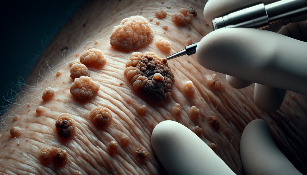
Seborrheic keratosis is a common benign (noncancerous) skin growth that tends to appear in middle age and becomes more prevalent as individuals get older. These slow-growing lesions are usually brown, black, or light tan in color, with a waxy or scaly appearance and a slightly raised texture. They are harmless, not contagious, and do not require treatment unless they become irritated or are cosmetically undesirable.
Definition
Seborrheic keratosis occurs when skin cells, known as keratinocytes, multiply rapidly, resulting in a non-cancerous growth. This condition has a likely genetic component, with certain mutations linked to its development. Sun exposure and changes in estrogen levels have also been associated with seborrheic keratosis.
RELATED: Melanoma: Early Detection and Treatment Options Explained
Appearance
Seborrheic keratoses can vary in size, ranging from very small to more than 1 inch (2.5 centimeters) across. They may appear as a single growth or in multiple clusters. The growths can be round or oval-shaped, with a waxy or rough surface texture. Some may resemble warts, while others have a characteristic “pasted on” look. The color of seborrheic keratoses can range from light tan to brown or black.
Common locations
Seborrheic keratoses can develop anywhere on the body, but they most commonly appear on sun-exposed areas such as the face, neck, chest, or back. In individuals with Black or brown skin, very small growths may cluster around the eyes or elsewhere on the face, a condition known as dermatosis papulosa nigra.
Causes and Risk Factors
The exact cause of seborrheic keratosis remains unknown. However, several factors have been associated with an increased risk of developing this condition:
- Age: Seborrheic keratosis is more common in older individuals, with the condition rarely affecting children, adolescents, and young adults. The suggestion is that chronic exposure to ongoing stimuli over time may contribute to the development of seborrheic keratosis.
- Genetics: Individuals with a family history of seborrheic keratosis are more likely to be affected than those without a family history. This increased risk is likely due to a genetic susceptibility passed down from parents to children. Specific gene mutations, such as those in the FGFR3, PIK3CA, RAS, AKT1, and EGFR genes, have been linked to individuals with seborrheic keratosis.
- Sun exposure: Some research suggests that frequent sun exposure may play a role in causing seborrheic keratosis. Eruptive seborrheic keratosis has been noted to often follow an episode of sunburn. However, the mechanism by which sunlight causes these growths remains unclear, particularly since they can appear on both sun-exposed and covered skin.
- Hormonal changes: Women sometimes notice the appearance of seborrheic keratosis during periods of significant hormonal changes, such as during pregnancy or after starting estrogen replacement therapy.
Other factors that may be involved in causing seborrheic keratosis include friction in skin folds during movement and the presence of certain skin conditions like dermatitis, with the growths more likely to appear following a flare-up. It is important to note that seborrheic keratosis is not contagious and does not spread to other parts of the body or other people. Additionally, while some have suggested that viruses like human papillomavirus may play a role, this appears unlikely. Seborrheic keratosis also does not seem to arise from mutations in tumor suppressor genes.
Diagnosis and Differentiation
Seborrheic keratosis has a characteristic “stuck on” appearance that allows experienced clinicians to easily identify the condition. However, due to its clinical heterogeneity, it can mimic other malignant skin disorders. A thorough evaluation is necessary to accurately diagnose seborrheic keratosis and rule out skin cancer.
Clinical examination
The diagnosis of seborrheic keratosis is typically made through clinical observation. These lesions present as raised, scaly, brown-to-black papules firmly attached to the skin. If there is uncertainty or concern for malignancy, such as ulcerated lesions, rapid change in size, or very large lesions, a skin biopsy is recommended for confirmation.
Dermoscopy
Dermoscopy can aid in the clinical diagnosis of seborrheic keratosis. Common dermoscopic findings include:
- Comedo-like openings
- Milia-like cysts
- Gyri and sulci
- Hairpin vessels
Features of melanocytic lesions, like pigment networks or globules, are absent in heavily pigmented seborrheic keratoses, though some typical dermoscopic features may be obscured.
RELATED: Leprosy (Hansen’s Disease): Symptoms, Causes, and Treatment Explained
Biopsy
A lesion biopsy is typically unnecessary for diagnosing seborrheic keratosis. However, in cases where the clinical or dermoscopic diagnosis is ambiguous, a biopsy and histopathologic examination are necessary to rule out melanoma, squamous cell carcinoma, or basal cell carcinoma.
Distinguishing from skin cancer
The differential diagnosis for seborrheic keratosis is broad and should include:
- Malignant melanoma
- Actinic keratosis
- Lentigo maligna
- Melanocytic nevus
- Squamous cell carcinoma
- Pigmented basal cell carcinoma
Overlapping lesions or high numbers of seborrheic keratosis can make the diagnosis and workup more difficult. Patients with numerous seborrheic keratoses require careful screening, as there is an increased chance of missing coexisting malignant lesions.
Although exceedingly uncommon, collision tumors and melanomas resembling seborrheic keratoses have been documented. The clinical and dermoscopy-based diagnosis of seborrheic keratosis-mimicking melanoma is challenging. Histologic and clinical examinations are crucial to accurately diagnose lesions that exhibit inconclusive clinical and dermoscopic characteristics.
Treatment Options
Although seborrheic keratosis is a benign condition that does not require treatment, many patients opt for removal due to cosmetic concerns or irritation. Several treatment modalities are available, including cryotherapy, electrocautery, curettage, laser therapy, and topical treatments.
Cryotherapy
Cryotherapy, the most common treatment for seborrheic keratosis, involves freezing the growth with liquid nitrogen using a cotton swab or spray gun. This method effectively destroys the lesion, causing it to fall off within days. Thicker lesions may require multiple freeze-thaw cycles. Cryotherapy is generally well-tolerated but can cause erythema, pain, and bulla formation.
Electrocautery
Electrocautery, also known as electrodesiccation, uses an electric current to destroy the seborrheic keratosis. This method is often combined with curettage and requires local anesthesia. The procedure involves scraping the lesion with a curette followed by electrodesiccation using a hyfrecator or cautery unit. This process is typically repeated multiple times to ensure complete removal. Electrodesiccation with curettage provides efficient results with a low complication rate, although scarring and hyperpigmentation may occur.
Curettage
Curettage involves scraping the seborrheic keratosis using a curette, a spoon-shaped surgical instrument. This method is often used in combination with electrodesiccation and requires local anesthesia. Curettage alone may be sufficient for superficial, epidermal lesions without dermal invasion.
Laser therapy
Laser therapy, including ablative (CO2 and erbium:YAG) and non-ablative (potassium-titanyl phosphate and neodymium-doped:YAG) lasers, has shown efficacy in treating seborrheic keratosis. The 532 nm picosecond laser has demonstrated success in treating seborrheic keratosis in Asian patients with minimal adverse effects. Laser therapy can provide precise removal of the lesion with reduced risk of damaging surrounding tissue.
RELATED: Preventing Lyme Disease: Symptoms, Causes, and Treatment Guide
Topical treatments
Various topical agents have been studied for the treatment of seborrheic keratosis, including:
- Alpha-hydroxy acids
- Urea ointment
- Vitamin D analogs (tacalcitol and calcipotriol)
- Diclofenac gel
- Potassium dobesilate
- 40% hydrogen peroxide solution
These topical medications work through different mechanisms, such as exfoliation, inflammation, or inhibition of tumor growth. While some studies have shown promising results, further research is needed to establish their efficacy and safety in treating seborrheic keratosis.
Conclusion
Seborrheic keratosis, while generally harmless, can have a significant impact on a person’s appearance and peace of mind. Understanding its causes, how to tell it apart from more serious conditions, and the available treatment options gives people the knowledge to handle this common skin issue. This awareness helps individuals make informed decisions about whether to seek treatment and which method might work best for them.
In the end, managing seborrheic keratosis is about balancing medical needs with personal preferences. While these growths don’t usually need treatment for health reasons, many choose to remove them to improve their appearance or ease discomfort. With various treatment options available, from freezing to laser therapy, there’s likely a solution that fits each person’s unique situation. As with any skin concern, it’s always a good idea to chat with a dermatologist to figure out the best approach.

