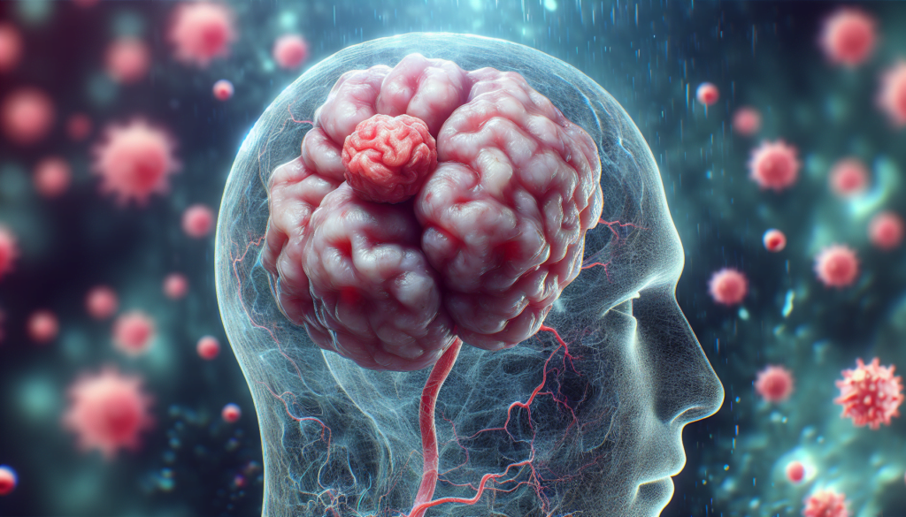
Astrocytoma, a term that denotes a group of brain tumors, stands as a critical subject within the medical field due to its complexity and the imperative need for effective management. Understanding what is astrocytoma, including its various types and the nuances behind its causes, is essential for both medical practitioners and patients navigating this challenging diagnosis. The significance of astrocytoma lies not only in its potential impact on patient well-being and survival but also in the insights it provides into the broader landscape of brain cancer research and treatment approaches. The ability to accurately diagnose and effectively treat astrocytoma has far-reaching implications, underscoring the importance of comprehensive knowledge about this condition.
This article aims to serve as a thorough guide, delving into the essential aspects of astrocytoma, from its symptoms and causes to the array of treatment options available. A closer look at astrocytoma symptoms helps to shed light on the early warning signs that may prompt further investigation. Additionally, understanding astrocytoma causes and risk factors plays a pivotal role in both prevention and early detection strategies. The section on diagnosis and tests will navigate through the methodologies employed to accurately identify astrocytoma, while the exploration of astrocytoma treatment outlines the standard and emerging therapies in the fight against this type of brain tumor. Furthermore, insights into prognosis and life expectancy will offer a glimpse into the outcomes associated with different astrocytoma types, enhancing the overall comprehension of this complex condition.
What is Astrocytoma?
Definition and Origin
Astrocytoma is a type of cancer that occurs in the brain or spinal cord. It originates in cells known as astrocytes, which are star-shaped glial cells located in the cerebrum. These cells play a crucial role in supporting and connecting nerve cells within the brain and spinal cord. Astrocytomas can manifest as either benign (noncancerous) or malignant (cancerous) tumors, with their growth rates and potential to infiltrate nearby tissues varying significantly.
Astrocytes, the cells from which these tumors derive, are part of the brain’s supportive tissue. Astrocytomas represent the most common form of glioma, a broader category of tumors that arise from glial cells. These tumors are most prevalent in individuals over the age of 45 and are slightly more common in men than women.
Types of Astrocytoma
Astrocytomas are classified into several types, each varying in terms of malignancy, growth rate, and typical age of onset:
- Pilocytic Astrocytoma (Grade I): This type is predominantly found in children and teenagers and is known for its slow growth. It is generally considered benign and has a favorable prognosis when surgically removed.
- Diffuse Astrocytoma (Grade II): Often referred to as low-grade astrocytoma, this type grows slowly and is usually seen in adults between the ages of 20 and 50. Although it can infiltrate nearby tissues, it is less aggressive than higher-grade tumors.
- Anaplastic Astrocytoma (Grade III): This is a more aggressive form that spreads quickly and is challenging to remove surgically due to its deep-rooted growth into surrounding brain tissue. It most commonly affects individuals between 30 and 50 years old.
- Glioblastoma (Grade IV): The most severe form of astrocytoma, glioblastoma is highly malignant and known for its rapid growth and poor prognosis. It typically occurs in adults aged 50 to 70 and can develop in various brain regions, including the spinal cord and brain stem.
Other notable types include the pineal astrocytic tumor, which affects the pineal gland, and brain stem gliomas, which are rare high-grade astrocytomas primarily affecting children.
Astrocytomas are graded by the World Health Organization (WHO) based on their growth rate and potential to spread to adjacent brain tissue, with Grade I being the least aggressive and Grade IV being the most aggressive. This classification helps guide treatment decisions and provides insight into the likely prognosis for affected individuals.
Symptoms of Astrocytoma
Common Symptoms
Astrocytomas present a range of symptoms that can significantly impact the quality of life and functionality of affected individuals. Common symptoms include persistent headaches, often more severe in the morning or causing awakening from sleep, which may indicate increased intracranial pressure. Patients may also experience nausea and vomiting, fatigue, and seizures, which are disruptive and can be alarming.
Cognitive and sensory impairments are also prevalent, with many experiencing decreased cognitive abilities, memory loss, and mood changes such as depression. Vision problems, including double or blurred vision, and speech difficulties are common, potentially affecting communication and daily activities.
Motor function can be compromised, manifesting as limb weakness, abnormal reflexes, or grasp difficulties. These symptoms collectively contribute to a decreased ability to perform daily tasks and maintain independence.
Symptoms Based on Tumor Location
The location of an astrocytoma within the brain or spinal cord greatly influences the specific symptoms experienced by the patient. Tumors in the brain can lead to personality changes, altered mental status such as delirium or dementia, and specific cognitive issues like aphasia or trouble speaking. These effects can alter a person’s social interactions and personal behavior significantly.
When astrocytomas develop in the spinal cord, they primarily cause physical symptoms such as weakness and disability in the affected areas, which can lead to challenges in mobility and physical function. The growth rate of the tumor also plays a critical role in symptom manifestation. Lower grade astrocytomas, often larger in size before they become symptomatic, tend to displace rather than destroy brain tissue and are associated with less swelling. In contrast, higher grade, malignant astrocytomas grow more rapidly and are more destructive, leading to quicker onset and more severe symptoms.
Causes and Risk Factors
Astrocytomas, like many other forms of cancer, arise from a complex interplay of genetic and environmental factors. Understanding these factors is crucial for assessing risk and developing preventive strategies.
Genetic Factors
Astrocytomas can sometimes occur as part of hereditary syndromes caused by inherited DNA mutations. These genetic conditions increase the likelihood of developing astrocytomas alongside other types of tumors:
- Li-Fraumeni Syndrome: This condition results from mutations in the tumor suppressor gene p53. It is characterized by the early onset of multiple cancers, including astrocytomas, breast cancer, and bone cancers.
- Turcot Syndrome: Associated with mutations in several tumor suppressor genes, including APC and MMR, this syndrome typically presents with early onset of colon cancer and astrocytomas.
- Neurofibromatosis Type 1: Caused by mutations in the NF1 gene, this disorder leads to the early development of astrocytomas, peripheral nerve tumors, and distinctive skin changes.
- Tuberous Sclerosis: This rare genetic disorder is often linked with mental retardation and the early development of subependymal giant cell astrocytoma (SEGA).
These syndromes highlight the significant role that genetics can play in the development of astrocytomas. Additionally, it has been observed that brain tumors, including astrocytomas, can cluster within families, suggesting a hereditary component beyond these well-defined syndromes.
Environmental Factors
Environmental exposures also contribute to the risk of developing astrocytomas, although these factors are generally less well-defined:
- Ionizing Radiation: Exposure to ionizing radiation, particularly during childhood as part of therapeutic treatment for other cancers, has been linked with a delayed onset of astrocytomas. The risk increases with the dose of radiation received and the young age at exposure.
- Chemical Exposure: There are suspicions, though not conclusively proven, that exposure to certain chemicals may increase the risk of astrocytomas. For example, veterans exposed to Agent Orange during the Vietnam War may have a higher risk of developing these tumors.
- Cellular Phones: Despite ongoing debates and research, there is currently no conclusive evidence to suggest that the use of cellular phones contributes to the development of astrocytomas.
In summary, while many cases of astrocytoma appear to occur sporadically, a combination of genetic predispositions and environmental exposures can significantly influence the risk. These factors are critical in understanding the complex etiology of astrocytomas and guiding both preventive and therapeutic strategies.
Diagnosis and Tests
Initial Examination
The diagnostic process for astrocytoma begins with a thorough initial examination. This includes a detailed review of the patient’s medical and family history, focusing on any past illnesses that may have compromised the immune system or involved radiation therapy. The patient’s habits and lifestyle are also discussed. A neurological exam is conducted to assess changes in vision, hearing, balance, coordination, strength, and reflexes. These observations help pinpoint which part of the brain might be affected by the tumor.
Imaging Tests
Imaging tests play a crucial role in diagnosing astrocytoma by providing detailed pictures of the brain. The primary imaging technique used is Magnetic Resonance Imaging (MRI), which utilizes radio waves and magnets to create detailed images of brain structures. During an MRI, a contrast dye may be injected to highlight the tumor’s location. Specialized MRI techniques such as functional MRI, perfusion MRI, and tractography are employed to map brain activity, identify areas with reduced blood flow, and visualize white matter tracts, respectively.
For patients unable to undergo MRI due to implants like pacemakers or cochlear implants, Computed Tomography (CT) scans are used. These scans combine multiple X-rays to construct comprehensive images of the brain’s structure. Additional imaging options include positron emission tomography (PET) scans, which use a small amount of radioactive glucose to detect tumor growth.
Biopsy
A biopsy is a definitive method for diagnosing astrocytoma. It involves the removal of a small tissue sample from the tumor, which is then examined under a microscope to confirm the presence of cancer cells. This procedure helps determine the tumor’s grade, indicating its aggressiveness. In cases where the tumor is difficult to access, a needle biopsy may be performed. Advanced techniques like stereotactic biopsy utilize a computerized navigation system to accurately target the tumor during tissue removal. This procedure is often integrated with molecular DNA testing to provide a precise diagnosis and guide treatment planning.
Treatment Options
Surgery
Surgery is often the initial treatment for astrocytomas and aims to remove as much of the tumor as possible. The extent of tumor resection can significantly impact subsequent treatment options and overall prognosis. Advanced surgical techniques such as neuronavigation, awake surgery, and motor mapping during general anesthesia enhance the precision and safety of the procedure, minimizing damage to surrounding healthy brain tissue. In cases where complete removal is not possible, reducing the tumor’s size can alleviate symptoms and improve the efficacy of other treatments like radiation and chemotherapy.
Radiation Therapy
Radiation therapy is a cornerstone in the management of astrocytoma, particularly for high-grade tumors. It involves the use of high-energy beams to target and destroy cancer cells. Techniques such as external beam radiation therapy, intensity-modulated radiation therapy, and image-guided radiation therapy are employed to maximize the dose to the tumor while protecting surrounding healthy tissue. Radiation is typically administered daily over several weeks following surgery. For tumors that recur, options like Gamma Knife radiosurgery may be used to enhance the effects of radiation.
Chemotherapy
Chemotherapy involves the use of drugs to kill cancer cells and is a standard treatment following surgery, especially for high-grade astrocytomas. The treatment regimen usually involves cycles of administration, allowing periods of recovery.
Targeted Therapy
Targeted therapies offer a more precise approach to treatment by focusing on specific molecular targets associated with cancer growth. Newer therapies, including peptide-based treatments, are being explored for their potential to deliver drugs directly to tumor cells, minimizing effects on healthy tissue. These therapies are often used in combination with conventional treatments to enhance efficacy and reduce side effects.
Prognosis and Life Expectancy
Astrocytoma prognosis varies widely based on several factors, including the tumor’s grade, the patient’s overall health, and the treatments applied. Understanding these variables is crucial for both patients and medical professionals when discussing life expectancy and managing expectations.
Survival Rates by Grade
The survival rates for astrocytoma differ significantly according to the tumor’s grade. Here are the American Cancer Society’s reported five-year survival rates for various grades of astrocytoma as of 2023:
- Pilocytic Astrocytoma (Grade I): The most favorable prognosis among astrocytomas, with a five-year survival rate of 97% for children and teenagers.
- Diffuse Astrocytoma (Grade II): This low-grade tumor has a five-year survival rate ranging from 73% for patients aged 20 to 44, to 26% for those aged 55 to 64.
- Anaplastic Astrocytoma (Grade III): A more aggressive form with a five-year survival rate of 58% for patients aged 20 to 44, decreasing to 15% for those aged 55 to 64.
- Glioblastoma (Grade IV): The most aggressive form with poor prognosis, showing a five-year survival rate of 22% for patients aged 20 to 44, and only 6% for those aged 55 to 64.
These statistics illustrate the marked variation in survival based on tumor grade and age at diagnosis.
Factors Affecting Prognosis
Several key factors influence the prognosis of individuals diagnosed with astrocytoma:
- Age and Overall Health: Younger patients generally have better outcomes. For instance, the survival rates are notably higher in younger age groups across all tumor grades.
- Tumor Characteristics: Factors such as the size, location, and whether the tumor has spread significantly affect prognosis. Tumors in critical areas like the brainstem are associated with poorer outcomes due to their impact on vital functions.
- Treatment Received: The extent of surgery, the use of radiation therapy, and chemotherapy play pivotal roles. Patients who undergo more complete tumor resections generally have better survival rates.
- Genetic Factors: Conditions like Li-Fraumeni Syndrome and Neurofibromatosis Type 1 can predispose individuals to poorer outcomes due to the aggressive nature of the associated astrocytomas.
Lifestyle factors also contribute to overall health and can influence cancer progression and recovery. Maintaining a healthy diet, regular exercise, avoiding tobacco, and managing stress are recommended for improving quality of life and potentially enhancing survival rates.
As research progresses and treatment approaches evolve, there is hope that survival rates for astrocytoma will continue to improve, offering patients better prospects than historical data might suggest.
Conclusion
Throughout this comprehensive guide, we explored the intricate nature of astrocytoma, delving into its symptoms, causes, diagnosis processes, and the multifaceted approaches to treatment. Our journey illuminated not only the challenges posed by this type of brain tumor but also the advancements in medical research and treatment options that offer hope to those affected. By understanding the gradation of astrocytomas and their manifestations, we’ve equipped readers with the knowledge necessary to navigate the complexities of diagnosis and treatment, reinforcing the article’s core message of enlightenment and empowerment for patients and their families.
The significance of ongoing research and the potential for new discoveries cannot be overstated, as they hold the promise of improving prognosis and life expectancy for individuals with astrocytoma. As we look toward the future, it’s clear that a multidisciplinary approach involving surgery, radiation therapy, chemotherapy, and targeted therapies plays a crucial role in optimizing patient outcomes. This guide serves as a testament to the resilience of those battling astrocytoma and underscores the importance of continuous learning, advocacy, and support within the medical community and beyond.

