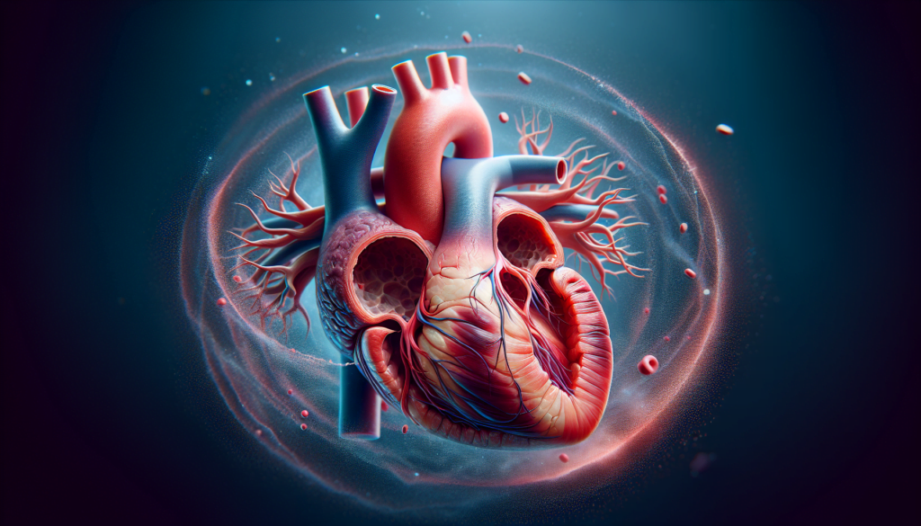
An atrial septal defect (ASD) is an often-overlooked heart condition that can have significant impacts if undiagnosed and untreated. Marked by an opening in the atrial septum, the wall that separates the heart’s upper chambers, this congenital defect allows oxygen-rich blood to mix with oxygen-poor blood. Understanding the nuances of atrial septal defect symptoms, causes, and treatments is crucial for timely intervention and management. Highlighting the importance of this condition underscores the need for awareness and comprehensive care for those affected, offering a clearer perspective on a condition that affects a notable number of individuals globally.
This article delves into the types of atrial septal defect, identifying specific atrial septal defect causes and risk factors that predispose individuals to this condition. Armed with knowledge, the reader will learn to recognize the signs and symptoms unique to ASD, facilitating early detection. Comprehensive coverage on the latest in diagnosis and testing paves the way for an informed understanding of atrial septal defect treatment options. From traditional surgical techniques to cutting-edge non-invasive procedures, the exploration of treatment pathways promises to illuminate the advancements in managing and possibly rectifying this heart condition, providing a beacon of hope for affected individuals and their families.
Understanding Atrial Septal Defect (ASD)
Definition and Basic Overview
An atrial septal defect (ASD) is a congenital heart defect characterized by a hole in the atrial septum, the wall that divides the heart’s two upper chambers, the atria. This condition is present at birth and can vary significantly in size. Small ASDs may never cause problems and can close on their own during infancy or early childhood. However, large or long-standing ASDs can lead to serious complications, necessitating medical intervention to prevent damage to the heart and lungs.
ASD occurs when the septum, which is supposed to form a solid wall between the atria, fails to develop properly, leaving an opening. This defect allows oxygen-rich blood from the left atrium to mix with oxygen-poor blood in the right atrium. Depending on the size of the hole, this can lead to an increased volume of blood passing through the lungs, which can strain both the heart and the lungs over time.
Importance of Early Detection
Early detection of an atrial septal defect is crucial due to the potential for significant health complications if left untreated. Serious congenital heart defects like ASD are often identified shortly after birth or during early childhood. Recognizing the symptoms early can lead to timely and effective treatment, significantly improving the prognosis for affected individuals.
Symptoms prompting immediate medical consultation include trouble breathing, especially in children, and signs such as shortness of breath during activity, unusual tiredness, swelling in the legs, feet, or abdominal area, and irregular heartbeats. These symptoms can indicate an increase in the workload on the heart and lungs caused by the abnormal blood flow through the ASD.
For large defects that do not close on their own, medical interventions such as percutaneous procedures or surgery might be required. These treatments prevent further complications such as stroke, heart rhythm problems (dysrhythmias), and pulmonary hypertension, which can lead to increased mortality if unaddressed.
In conclusion, understanding the basic mechanics and implications of an atrial septal defect, coupled with the importance of early detection, equips individuals and healthcare providers with the knowledge necessary to manage this condition effectively. Early intervention not only helps in mitigating the immediate health risks but also reduces the long-term impact on an individual’s quality of life.
Types of Atrial Septal Defects
Atrial Septal Defects (ASDs) are classified into several types based on their location and the specific structural characteristics of the atrial septum. Understanding these types is crucial for accurate diagnosis and appropriate treatment planning.
Secundum ASDs
Secundum ASDs are the most prevalent type, constituting approximately 75% of all atrial septal defects. They occur in the middle of the atrial septum, known as the fossa ovalis. Typically, these defects are associated with a left-to-right shunt of blood, which can lead to volume overload on the right side of the heart. The magnitude of blood flow through a Secundum ASD depends on the size of the defect and the relative pressures in the atria. Although many cases remain asymptomatic into adulthood, significant shunts can lead to symptoms such as fatigue and respiratory distress. Treatment options include percutaneous device closure and surgical repair, which are highly effective with minimal risk.
Primum ASDs
Primum ASDs are located at the lower part of the atrial septum, near the atrioventricular valves. These defects are less common and often occur in conjunction with other congenital heart anomalies, such as endocardial cushion defects. Primum ASDs are particularly associated with conditions like Down Syndrome. The defect allows a mixing of oxygen-rich and oxygen-poor blood, leading to similar hemodynamic consequences as other ASDs but often requires a more complex intervention due to the associated anomalies. Diagnosis is typically made through transthoracic echocardiography, which can reveal the defect and associated abnormalities in heart structure.
Sinus Venosus ASDs
Sinus Venosus ASDs are rare and occur at the junction of the superior or inferior vena cava with the right atrium, away from the central part of the atrial septum. These defects are often associated with anomalous pulmonary venous return, where one or more pulmonary veins drain into the right atrium instead of the left, exacerbating the shunt’s effects. The clinical manifestations may not be apparent in childhood but can lead to significant symptoms like dyspnea on exertion and arrhythmias as the patient ages. Advanced imaging techniques such as transesophageal echocardiography (TEE) or cardiac MRI are often required for accurate diagnosis due to the defect’s posterior location.
Coronary Sinus Defects
Coronary Sinus Defects, also known as unroofed coronary sinus syndrome, are the least common type of ASD. These involve the absence of part of the wall that separates the coronary sinus (which carries blood from the myocardium to the right atrium) from the left atrium. This defect creates a left-to-right shunt at the atrial level. It is frequently associated with a persistent left superior vena cava. The defect may not cause noticeable symptoms in early life but can lead to complications like right heart overload and arrhythmias over time. Diagnosis often requires a high index of suspicion and detailed imaging studies. Treatment typically involves surgical correction to prevent long-term complications.
Each type of atrial septal defect presents unique challenges in both diagnosis and management, emphasizing the need for a thorough understanding of their distinct characteristics.
Causes and Risk Factors
Genetic Factors
Atrial Septal Defect (ASD) can be influenced by genetic factors, with several inherited mutations playing a crucial role in the development of this condition. Mutations in genes encoding cardiac transcription factors and sarcomeric proteins have been identified as underlying causes for familial recurrence of non-syndromic congenital heart defects (CHD), particularly cardiac septal defects. Notably, the cardiac phenotypes most frequently seen in mutation carriers are ostium secundum atrial septal defects (ASDII).
Key transcription factors such as TBX5, NKX2-5, and GATA4 are fundamental in heart development, influencing processes like cardiac lineage determination, chamber formation, valvulogenesis, and septation. For instance, mutations in TBX5 result in Holt–Oram syndrome, characterized by malformations of the upper limb and ASD. Similarly, NKX2-5 mutations have been linked to ASD with atrioventricular conduction defects and are associated with increased risks of sudden cardiac death post-surgical repair due to conduction disturbances.
Furthermore, sarcomeric gene mutations, specifically in the MYH6 gene, have been identified in families with ASDII. These mutations highlight the genetic complexity and the varied phenotypic expressions of ASD, underlining the importance of genetic testing in families with a history of ASD to guide treatment options and preventive measures.
Environmental Influences
Environmental factors also play a significant role in the development of ASD. Several maternal and paternal risk factors have been identified, including obesity, advanced paternal age, and exposure to certain environmental toxins. Maternal factors such as smoking, alcohol consumption, and the use of specific medications during pregnancy, like antidepressants, have been shown to increase the risk of CHD in infants.
Exposure to environmental contaminants, many of which are classified as endocrine disruptors or teratogens, has been recognized as a significant risk factor. These substances can interfere with normal embryonic development either directly or indirectly by affecting placental function and altering the nutrient supply to the embryo. For instance, cigarette smoke contains multiple harmful substances that can cross the placenta, potentially leading to reduced oxygen and nutrient availability to fetal tissues.
Moreover, studies have shown a strong association between maternal obesity and an increased risk of ASD. Research indicates that obesity can significantly elevate the incidence rate of ASD, underscoring the need for public health interventions aimed at managing obesity to potentially reduce the prevalence of this congenital defect.
In conclusion, both genetic and environmental factors contribute to the risk and development of atrial septal defects. Understanding these factors is crucial for early detection, prevention, and management of ASD, ultimately improving outcomes for affected individuals.
Signs and Symptoms
Identifying Common Symptoms
Atrial septal defect (ASD) is a congenital heart defect that may not always produce noticeable symptoms, especially if the defect is small. However, when symptoms do manifest, they can vary depending on the age of the individual and the size of the defect. Common symptoms include:
- Shortness of Breath: This is often more noticeable during exercise or physical activity, but can also occur during rest in more severe cases.
- Tiredness: Individuals with ASD may experience fatigue, particularly after physical activity.
- Swelling: Swelling of the legs, feet, or abdominal area can occur due to fluid accumulation, a condition often exacerbated by the increased blood flow to the lungs that ASD can cause.
- Irregular Heartbeats: Also known as arrhythmias, these can range from skipped heartbeats to sensations of a quick, pounding, or fluttering heartbeat, known as palpitations.
In children, the symptoms might not be pronounced and are often detected during routine medical checks. A heart murmur, typically discovered by a healthcare provider during a physical examination, is frequently the first indicator of ASD. Other possible signs in children include poor appetite, delayed growth, and recurrent respiratory infections.
When to Seek Medical Attention
Immediate medical attention should be sought if severe symptoms present themselves, especially in children. Key indicators for urgent care include:
- Trouble Breathing: If a child exhibits difficulty breathing, it is critical to seek emergency medical help.
- Severe Swelling and Fatigue: These symptoms can indicate a significant compromise in heart function due to the defect.
For adults, symptoms might not appear until later in life, often by the age of 40. Adults should consult healthcare professionals if they experience:
- Persistent Shortness of Breath: Especially if it occurs during activities that were previously manageable without difficulty.
- Unusual Heart Rhythms: Any changes in heartbeat, such as palpitations or feeling a fast heartbeat, should prompt a consultation with a healthcare provider.
It is essential for anyone with suspected or known ASD to be aware of these symptoms and to have regular follow-ups with a healthcare provider. Early detection and treatment can significantly improve the quality of life and prognosis for individuals with atrial septal defects.
Diagnosis and Testing
Diagnostic Methods
Atrial Septal Defect (ASD) diagnosis involves a combination of physical examinations and advanced imaging techniques to accurately assess the presence and severity of the defect. The diagnostic process typically begins with the detection of a heart murmur during a physical examination using a stethoscope. This whooshing sound may indicate abnormal blood flow within the heart.
Echocardiogram
The primary tool for diagnosing ASD is the echocardiogram, which uses sound waves to create detailed images of the heart’s structure and function. This test can show the size and location of the defect, the condition of the heart chambers and valves, and how blood moves through the heart.
Electrocardiogram (ECG or EKG)
An ECG records the electrical activity of the heart and helps in detecting arrhythmias and other heart function irregularities that might be influenced by ASD.
Chest X-ray
A chest X-ray provides images of the heart and lungs and can indicate changes such as an enlargement of the right atrium or right ventricle, which are common in ASD cases.
Advanced Imaging Techniques
For more complex cases or when initial tests are inconclusive, advanced imaging techniques such as Cardiac MRI and CT scans are employed. These tests offer detailed views of the heart’s structure and are particularly useful for diagnosing less common forms of ASD or associated defects.
Specialized Echocardiography
Transthoracic echocardiography (TTE) is often supplemented by Transesophageal echocardiography (TEE) and Intracardiac echocardiography (ICE), especially during surgical or percutaneous repair procedures. TEE provides detailed images of the heart from the esophagus, which is particularly useful for viewing the upper part of the heart and the atrial septum. ICE involves the insertion of a camera directly into the heart through a vein, offering a close-up view of the defect.
Interpreting Test Results
The interpretation of diagnostic test results is crucial for determining the appropriate management strategy for ASD. Here are key aspects involved in interpreting these results:
Assessing the Size and Impact of ASD
The size of the atrial septal defect and the extent of left-to-right blood shunting are critical factors assessed during diagnosis. Tests like echocardiography with Doppler flow studies help in measuring the shunt’s size and predicting its physiological impact.
Evaluating Heart Function
ECG and echocardiogram results are analyzed to evaluate the overall function of the heart, focusing on potential complications like arrhythmias and heart chamber enlargement.
Identifying Associated Conditions
Advanced imaging tests such as MRI and CT scans not only confirm the presence of ASD but also identify any associated abnormalities or complications like pulmonary hypertension or structural heart changes.
Continuous Monitoring
For some patients, particularly those with minimal symptoms or small defects, ongoing monitoring through regular echocardiography might be recommended to observe any changes in the condition over time.
The comprehensive evaluation and interpretation of these tests ensure that each individual with ASD receives a tailored treatment plan that addresses both the defect and any related health issues, aiming for optimal long-term outcomes.
Treatment Options and Management
Non-surgical Approaches
The management of atrial septal defect (ASD) varies based on the size of the defect and the presence of symptoms. Small ASDs that do not close naturally during childhood may not require surgical intervention. Instead, regular health checkups are essential to monitor the condition. Medications do not repair the defect but can alleviate symptoms. Commonly prescribed medications include:
- Beta blockers: These are used to control heart rate and prevent arrhythmias.
- Anticoagulants: Also known as blood thinners, these medications help reduce the risk of blood clots, which is crucial in preventing stroke or other complications.
- Diuretics: These help reduce fluid buildup in the body, including the lungs, thus easing the strain on the heart.
It is important to note that while these medications help manage symptoms, they do not correct the underlying defect.
Surgical Interventions
For medium to large ASDs, or in cases where the defect leads to significant symptoms or complications, surgical intervention may be necessary. The two primary methods for repairing ASDs are:
- Catheter-Based Repair:
- This procedure is typically used for Secundum ASDs, which are the most common type.
- A catheter is inserted into a vein, usually in the groin, and guided to the heart.
- A mesh patch or plug is deployed through the catheter to close the hole. Over time, heart tissue grows over this patch, effectively sealing the defect permanently.
- Open-Heart Surgery:
- This approach is required for other types of ASDs, such as Primum, Sinus Venosus, and Coronary Sinus ASDs.
- It involves a chest incision to access the heart, where patches are used to close the defect.
Minimally invasive techniques, including robot-assisted surgery, may be employed to reduce recovery times and improve outcomes. These methods use smaller incisions and are generally less traumatic than traditional open-heart surgery.
Post-treatment Care
Following ASD closure, whether by surgical or catheter-based methods, ongoing monitoring is crucial. Regular imaging tests and health checkups help ensure that the heart and lungs are functioning well and that no new complications arise. Key aspects of post-treatment care include:
- Monitoring for Arrhythmias: Irregular heartbeats are a potential complication after ASD repair.
- Watching for Pulmonary Hypertension: This serious condition can develop if the ASD was large or if the repair was performed later in life.
- Lifestyle Adjustments: Patients may need to modify their activities temporarily to allow the heart to heal.
Patients are typically advised to avoid strenuous activities for a period, depending on the type of procedure performed. For those who underwent catheter-based repair, normal activities can often be resumed within a week, while more time may be needed after open-heart surgery.
It is essential for patients to attend all follow-up appointments and adhere to their healthcare provider’s recommendations to optimize recovery and prevent future complications.
Conclusion
Through exploring the intricacies of atrial septal defects, from their symptoms and diagnostic procedures to the treatment methodologies available, it becomes clear how vital knowledge and early intervention are in managing this congenital heart condition. The article underscored the importance of diagnosing ASD early to mitigate the potential for severe health complications, revealing a complex blend of genetic and environmental factors that contribute to its development. Equipped with this information, individuals can better understand the signs to watch for, ensuring timely medical consultation and treatment.
The exploration of treatment options, ranging from non-surgical approaches to advanced surgical interventions, highlighted significant advancements in medical practices aimed at improving the quality of life for those affected by ASD. As the medical field continues to evolve, the hope for those dealing with atrial septal defects shines brighter, underscored by the potential for successful management and minimization of long-term health impacts. It reinforces the piece’s core message: awareness, early detection, and proactive treatment are key in navigating the challenges posed by atrial septal defects, offering not just a chance at a healthier life, but also a testament to the strides made in congenital heart defect care.

