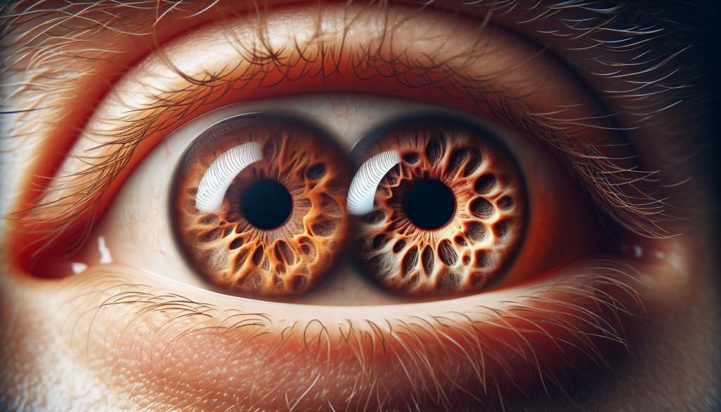
Anisocoria, a condition where the pupils are unequal in size, can be a sign of various underlying health issues. This eye abnormality occurs when one pupil dilates more or less than the other, resulting in a noticeable difference in appearance. While some cases of anisocoria are harmless and even considered normal, others may indicate serious medical conditions that require immediate attention.
Understanding the causes and treatments for anisocoria is crucial for proper diagnosis and management. This article delves into the symptoms associated with this condition, explores the diagnostic approach used by healthcare professionals, and discusses available treatment options. By shedding light on these aspects, readers will gain valuable insights into this intriguing eye condition and its potential implications for overall health.
Symptoms Associated with Anisocoria


Anisocoria can be accompanied by various symptoms depending on the underlying cause. Some individuals with anisocoria may not experience any additional symptoms, while others may have significant visual changes, discomfort, or neurological issues.
Visual changes
One of the most common symptoms associated with anisocoria is blurred vision. The difference in pupil size can cause an imbalance in the amount of light entering each eye, leading to difficulty focusing and reduced visual acuity. Additionally, some people with anisocoria may experience sensitivity to light (photophobia), particularly in the eye with the larger pupil. This can cause discomfort and make it challenging to function in bright environments.
RELATED: Bee Sting Relief: Expert Tips on Symptoms, Treatment, and Prevention
Eye pain
In some cases, anisocoria may be accompanied by eye pain or a feeling of pressure behind the affected eye. This pain can range from mild to severe and may be constant or intermittent. Eye pain associated with anisocoria can be a sign of a more serious underlying condition, such as glaucoma or inflammation, and should be evaluated by a healthcare professional.
Headache
Anisocoria can sometimes occur in conjunction with headaches, particularly in individuals with migraines. During a migraine episode, some people may experience temporary anisocoria along with other symptoms such as throbbing head pain, nausea, and sensitivity to light and sound. In rare cases, anisocoria may be a sign of a more severe headache disorder called ophthalmoplegic migraine, which can cause weakness or paralysis of the eye muscles.
Other neurological symptoms
Depending on the cause of anisocoria, individuals may experience additional neurological symptoms. These can include double vision (diplopia), drooping eyelid (ptosis), and weakness or numbness in the face or extremities. In some cases, anisocoria may be a sign of a serious neurological condition, such as a brain tumor, stroke, or aneurysm. If anisocoria is accompanied by any of these symptoms, it is crucial to seek immediate medical attention for proper diagnosis and treatment.
Diagnostic Approach to Anisocoria
When evaluating a patient with anisocoria, obtaining a thorough history is crucial. This includes determining the onset and duration of the anisocoria, any associated symptoms, and potential triggering factors such as medication use or recent eye trauma. Looking at old photographs can help establish if the anisocoria has been present for a long time.
History taking
During history taking, it is important to inquire about the use of eye drops, contact with substances like scopolamine patches or asthma inhalers, and any prior eye surgeries. A history of headaches, especially those associated with autonomic features, should be elicited as they may point towards conditions like Horner syndrome or trigeminal autonomic cephalalgias. Other relevant historical details include the presence of diplopia, ptosis, numbness, ataxia, dysarthria, or weakness, which can help localize the underlying pathology.
Physical examination
The physical examination should focus on accurately measuring pupil sizes in both light and dark conditions. Shining a light obliquely from below the patient’s face and using a handheld pupil gauge can aid in precise measurement. Pupil reactivity to light is typically graded on a scale of 0 (no reaction) to 4 (brisk reaction), with symmetry being more important than absolute values. The presence of dilation lag, characterized by the slow dilation of a miotic pupil in the dark, is a key feature of Horner syndrome.
RELATED: Understanding Bone Spurs (Osteophytes): Symptoms and Prevention Tips
Pupillary light reflex testing
Careful testing of the pupillary light reflex is essential. The swinging flashlight test can reveal a relative afferent pupillary defect (RAPD) in cases of unilateral optic nerve or severe retinal pathology. When performing this test, it is crucial to shine the light along the visual axis, which can be challenging in the presence of significant ocular misalignment.
Pharmacological tests
In some cases, pharmacological testing may be necessary to diagnose the cause of anisocoria. For instance, dilute pilocarpine (0.125%) can be used to differentiate Adie’s tonic pupil from other causes of pupillary dilation. In Horner syndrome, cocaine or apraclonidine eye drops can help confirm the diagnosis, while hydroxyamphetamine can aid in localizing the lesion to pre- or post-ganglionic neurons.
A comprehensive evaluation of anisocoria requires a systematic approach that includes a detailed history, careful physical examination, and appropriate diagnostic tests. By considering the various etiologies and localizing the underlying pathology, clinicians can effectively manage patients with this condition and prevent potential complications.
Treatment Options for Anisocoria
Addressing underlying causes
The treatment of anisocoria depends on identifying and addressing the underlying problem. For physiologic anisocoria, no treatment is needed. Mechanical anisocoria secondary to trauma may require surgery to correct the structural defect. Inflammatory conditions such as uveitis or acute angle closure glaucoma can be medically and/or surgically managed as indicated. Pharmacologic anisocoria typically resolves with cessation of the offending agent.
Symptomatic relief
If the patient experiences associated accommodation paresis, reading glasses or bifocals may be required. For those with mydriasis and glare, dilute pilocarpine, sunglasses, or FL-41 lenses may provide relief. Consultation with a neurologist or neuro-ophthalmologist is recommended for atypical cases, such as autoimmune autonomic ganglionopathy and trigeminal autonomic cephalalgias.
RELATED: Blister Prevention: Tips to Keep Your Skin Safe
Surgical interventions
Surgical care depends upon the specific etiology of anisocoria. Compressive third nerve palsies and aneurysms may necessitate neurosurgical intervention, while ophthalmologists can assist in other causes. Life-threatening conditions like stroke, aneurysm, hemorrhage, dissection, and tumor must be ruled out and/or managed appropriately to prevent potential complications.
Follow-up care
Follow-up, treatment, prognosis, and educational issues are contingent upon the underlying diagnosis of anisocoria. Patients should be educated that if they develop a sudden severe headache, blood in sputum, or a sudden blurring of vision with an associated anisocoria, they should seek medical attention promptly as these signs could indicate a serious issue that requires evaluation. Regular monitoring and follow-up with the appropriate specialists are crucial for the effective management of anisocoria and its associated conditions.
Conclusion
Anisocoria, with its various causes and treatments, has a significant impact on eye health and overall well-being. This condition, characterized by unequal pupil sizes, can range from harmless to potentially life-threatening, making proper diagnosis and management crucial. By understanding the symptoms, diagnostic approaches, and treatment options, individuals and healthcare providers can better address this eye abnormality and its underlying causes.
To wrap up, the comprehensive examination of anisocoria highlights the importance of timely medical attention and appropriate care. Whether it’s a benign physiological variation or a sign of a more serious condition, recognizing and addressing anisocoria can lead to improved outcomes. As research in this field continues, new insights and treatment methods may emerge, further enhancing our ability to manage this intriguing eye condition effectively.

