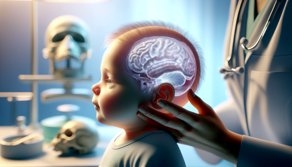
Craniosynostosis is a rare but serious condition that affects the growth and development of a baby’s skull. This disorder occurs when one or more of the fibrous joints between the bones of an infant’s skull close prematurely, causing the head to grow in an unusual shape. Early detection and treatment of craniosynostosis are crucial to prevent potential complications and ensure proper brain development.
Parents and healthcare providers need to be aware of the signs and symptoms of craniosynostosis to seek timely medical attention. This article will explore the key aspects of this condition, including its causes, common symptoms, diagnostic methods, and available treatment options. By understanding craniosynostosis better, families can make informed decisions and work with medical professionals to ensure the best possible outcomes for affected children.
Understanding Craniosynostosis
Definition and Types
Craniosynostosis is a condition in which one or more of the fibrous sutures in an infant’s skull prematurely fuses and turns into bone, changing the growth pattern of the skull. Craniosynostosis can be classified into different types based on the affected suture(s). Sagittal synostosis involves the sagittal suture that runs along the top of the head and is the most common type. Coronal craniosynostosis affects the coronal suture that extends from ear to ear and is the second most common. Metopic synostosis involves the metopic suture that runs from the top of the head down the middle of the forehead, while lambdoid craniosynostosis affects the lambdoid suture at the back of the head. Complex craniosynostosis is a term used when multiple cranial sutures are involved.
Causes and Risk Factors
The exact causes of craniosynostosis are not fully understood, but it is thought to involve a combination of genetic and environmental factors. In some cases, craniosynostosis is caused by certain genetic syndromes such as Apert syndrome, Crouzon syndrome, Pfeiffer syndrome, and Saethre-Chotzen syndrome. These syndromes are associated with mutations in specific genes involved in bone growth and development, such as FGFR1, FGFR2, FGFR3, TWIST1, and EFNB1. Non-syndromic craniosynostosis, which accounts for the majority of cases, is believed to have a multifactorial etiology involving both genetic predisposition and environmental influences. Studies have suggested that risk factors for non-syndromic craniosynostosis may include advanced parental age, maternal smoking, and certain medications used during pregnancy. Fetal constraint, such as intrauterine head compression, has also been proposed as a potential risk factor.
RELATED: Essential Facts About Rabies: Symptoms, Causes, and Vaccines
Prevalence and Demographics
Craniosynostosis occurs in approximately 1 in every 2,000 to 2,500 live births. The prevalence of different types of craniosynostosis varies, with sagittal synostosis being the most common, followed by coronal, metopic, and lambdoid synostosis. Complex craniosynostosis, involving multiple sutures, accounts for around 5% of all cases. Craniosynostosis affects males more frequently than females, with a male-to-female ratio of approximately 2:1. However, coronal craniosynostosis has a higher incidence in females. The condition is observed in all racial and ethnic groups, although some studies have suggested that there may be slight differences in the distribution of certain types of craniosynostosis among different populations. Early recognition and timely treatment of craniosynostosis are crucial to prevent potential complications and ensure optimal outcomes for affected infants. Healthcare providers and parents should be aware of the signs and symptoms associated with the different types of craniosynostosis, such as an abnormally shaped head, ridging along the affected sutures, and asymmetric facial features. Prompt referral to a multidisciplinary craniofacial team for further evaluation and management is essential to address the functional and aesthetic concerns associated with craniosynostosis.
Recognizing the Signs and Symptoms
Physical Characteristics
The most apparent sign of craniosynostosis is an abnormally shaped head. The specific head shape depends on which suture or sutures are affected. In sagittal synostosis, the head appears long and narrow, with a prominent forehead and possible ridging along the fused suture. Coronal synostosis results in a flattened forehead on the affected side and a raised eye socket, while the nose may deviate towards the unaffected side. Metopic synostosis leads to a triangular-shaped forehead when viewed from above, and the eyes may appear close together (hypotelorism). Lambdoid synostosis, although rare, causes flattening of the back of the head on the affected side, and one ear may be positioned lower than the other.
Developmental Concerns
While not all children with craniosynostosis experience developmental delays, some may face challenges in various domains. Delays in motor development, such as reaching milestones like crawling or walking, can be an early indicator of potential issues. Speech and language delays may also occur, with some children having difficulty in expressing themselves or understanding others. Cognitive development may be affected, with some children experiencing learning difficulties or struggles with attention and memory. It is essential for parents and healthcare providers to monitor the child’s development closely and address any concerns promptly.
RELATED: Personality Disorders: From Symptoms to Treatment Options
Associated Health Issues
In addition to the physical and developmental concerns, children with craniosynostosis may experience other health issues. Increased intracranial pressure (ICP) is a potential complication, especially in cases of multiple suture involvement or delayed treatment. Signs of increased ICP include irritability, vomiting, lethargy, and a bulging or tense fontanelle. Visual problems, such as strabismus (misaligned eyes) or papilledema (swelling of the optic nerve), can also occur due to increased pressure on the brain and eyes. Breathing difficulties, particularly in cases of midface hypoplasia (underdevelopment of the middle part of the face), may be present. Regular monitoring by a multidisciplinary team is crucial to identify and manage any associated health issues promptly. Early recognition of the signs and symptoms of craniosynostosis is crucial for timely intervention and optimal outcomes. Healthcare providers should educate parents about the characteristic physical features and potential developmental and health concerns associated with the condition. Routine well-child visits provide an opportunity for close monitoring of head growth and shape, as well as developmental milestones. If any concerns arise, prompt referral to a craniofacial specialist is essential for further evaluation and management. By increasing awareness and facilitating early detection, healthcare professionals can play a vital role in ensuring that children with craniosynostosis receive the care they need to thrive.
Diagnosis and Evaluation
Physical Examination
The diagnosis of craniosynostosis begins with a thorough physical examination of the infant’s head shape and facial features. Abnormalities in head shape, such as frontal bossing, cloverleaf skull, turricephaly, or dolichocephaly, can indicate specific types of craniosynostosis. Facial asymmetry, midface hypoplasia, and abnormal positioning of the ears may also be present. The examiner should carefully palpate the cranial sutures to assess for ridging or premature fusion. Associated features, such as syndactyly in Apert syndrome or the characteristic facial features of Crouzon syndrome, can provide clues to the underlying diagnosis.
Imaging Studies
Imaging plays a crucial role in confirming the diagnosis of craniosynostosis and guiding surgical planning. Plain radiographs, including anteroposterior and lateral views of the skull, can demonstrate characteristic findings such as bony bridging across the affected suture, sclerosis, and loss of suture clarity. However, the entire length of each suture may not be visible on plain films, and normal variations in skull shape can pose challenges in interpretation.
Computed tomography (CT) with three-dimensional (3D) reconstruction is considered the gold standard for diagnosing craniosynostosis. CT provides detailed visualization of the calvarial bones, sutures, and associated intracranial anomalies. Three-dimensional reconstructions allow for a comprehensive assessment of the craniofacial deformity and are invaluable for surgical planning. However, the radiation exposure associated with CT scans is a concern, particularly in young children who are more radiosensitive.
Ultrasound has emerged as a promising radiation-free alternative for evaluating cranial sutures in infants. High-frequency ultrasound can clearly demonstrate the patency or premature fusion of sutures in children younger than 12 months. It offers the advantage of bedside examination without the need for sedation. However, ultrasound is operator-dependent, and inexperienced personnel may miss the diagnosis.
Magnetic resonance imaging (MRI) is not routinely used for diagnosing craniosynostosis but can provide valuable information about associated brain abnormalities. Advanced MRI techniques, such as black bone imaging, have shown potential in visualizing cranial sutures and creating 3D reconstructions similar to CT scans.
Genetic Testing
Genetic testing is an important component of the diagnostic evaluation, particularly in cases of syndromic craniosynostosis. Identifying the underlying genetic mutation can guide genetic counseling, predict the risk of recurrence, and inform long-term management. Targeted gene sequencing panels or whole exome sequencing can detect mutations in genes commonly associated with craniosynostosis syndromes, such as FGFR2, FGFR3, TWIST1, and ERF. In some cases, parental testing may be recommended to determine if the mutation is de novo or inherited.
A multidisciplinary approach involving pediatricians, neurosurgeons, geneticists, and radiologists is essential for the comprehensive evaluation and management of infants with craniosynostosis. Early diagnosis through careful physical examination and appropriate imaging studies allows for timely intervention to prevent complications and optimize outcomes. Genetic testing provides valuable insights into the underlying etiology and guides long-term care. With advances in imaging techniques and genetic diagnostics, the diagnostic approach to craniosynostosis continues to evolve, enabling more precise and personalized management strategies.
Treatment Options and Management
Surgical Interventions
The primary goal of surgical treatment for craniosynostosis is to expand the cranial vault, providing adequate space for brain growth and development while simultaneously correcting the abnormal head shape. The specific surgical approach depends on the type and severity of craniosynostosis, as well as the age of the child at the time of intervention.
For single-suture craniosynostosis, such as sagittal or metopic synostosis, minimally invasive endoscopic techniques can be employed in infants younger than 3-4 months. These procedures involve small incisions, the use of an endoscope to visualize the affected suture, and removal of the fused suture to allow for brain expansion. Postoperatively, the infant typically wears a custom-fitted molding helmet to guide skull growth and achieve optimal head shape.
In older infants or those with more complex forms of craniosynostosis, open cranial vault remodeling procedures are often necessary. These surgeries involve larger incisions, removal of the affected bone, and reshaping of the skull to create a more normal head shape. The reshaped bone is then stabilized with resorbable plates and screws. Open procedures generally require longer hospital stays and have a higher risk of complications compared to minimally invasive techniques.
For syndromic craniosynostosis, such as Apert or Crouzon syndrome, multiple staged surgeries may be required to address both the cranial vault and midface abnormalities. These procedures are more complex and often involve a combination of techniques, such as distraction osteogenesis, to gradually advance the bones of the face and forehead.
RELATED: Effective Treatments for Peritonitis: What to Expect
Non-Surgical Approaches
In some cases of mild craniosynostosis, non-surgical interventions may be considered. Cranial molding helmets can be used to guide skull growth and improve head shape in infants with mild deformities or following endoscopic surgery. These custom-fitted helmets apply gentle pressure to specific areas of the skull, encouraging growth in the desired direction. Helmet therapy typically begins around 4-6 months of age and continues for several months, with regular adjustments to accommodate the child’s growth.
Physical therapy may also play a role in the management of craniosynostosis, particularly in cases where associated torticollis or developmental delays are present. Stretching exercises and positioning techniques can help improve neck range of motion and promote symmetrical head growth.
In rare instances, where the craniosynostosis is very mild and does not appear to be impacting brain development, a “watch and wait” approach may be taken, with close monitoring by the craniofacial team to ensure that no further intervention is necessary.
Post-Treatment Care and Follow-up
Following surgical correction of craniosynostosis, ongoing follow-up with the multidisciplinary craniofacial team is essential to monitor the child’s growth, development, and potential complications. Regular appointments with the neurosurgeon, plastic surgeon, and other specialists involved in the child’s care are typically scheduled at intervals ranging from a few weeks to several months, depending on the individual case.
During these follow-up visits, the team assesses the child’s head shape, growth, and overall development. Imaging studies, such as X-rays or CT scans, may be performed to evaluate the success of the surgical intervention and to identify any areas of concern. Neuropsychological evaluations may also be conducted to monitor the child’s cognitive development and identify any potential delays or learning difficulties.
If additional surgeries are required, such as in cases of syndromic craniosynostosis, the craniofacial team will coordinate the timing and planning of these procedures to optimize outcomes and minimize the number of interventions needed.
Long-term follow-up is crucial, as some children with craniosynostosis may experience ongoing challenges related to their condition, such as visual impairments, hearing loss, or developmental delays. By maintaining a close relationship with the craniofacial team, families can ensure that their child receives the necessary support and interventions to achieve the best possible outcomes.
Conclusion
Craniosynostosis has a significant impact on the growth and development of a child’s skull, making early detection and treatment crucial. Understanding the signs, symptoms, and available diagnostic methods empowers parents and healthcare providers to take swift action. The various treatment options, ranging from minimally invasive procedures to complex surgeries, offer hope for children affected by this condition. What’s more, ongoing research and advancements in surgical techniques continue to improve outcomes for patients.
To wrap up, managing craniosynostosis requires a team effort involving multiple specialists and long-term follow-up care. By raising awareness about this condition and promoting early intervention, we can help ensure that affected children receive the support they need to thrive. While challenges may arise, the combination of medical expertise, family involvement, and continued research paves the way for better outcomes and improved quality of life for those living with craniosynostosis.

