Have you ever experienced a painful lump behind your knee? You might be dealing with a Baker’s cyst, a common condition that can cause discomfort and limit mobility. This fluid-filled sac, also known as a popliteal cyst, forms when excess synovial fluid builds up in the back of the knee joint. Understanding the nature of a Baker’s cyst is crucial for those seeking relief and proper management.
This article delves into the world of Baker’s cysts, providing essential information to help you navigate this condition. We’ll explore its causes, symptoms, and diagnostic methods, as well as discuss various treatment options available. Whether you’re dealing with a Baker’s cyst yourself or simply want to learn more about this knee issue, this guide aims to give you a clear picture of what to expect and how to handle it effectively.
What is a Baker’s Cyst?
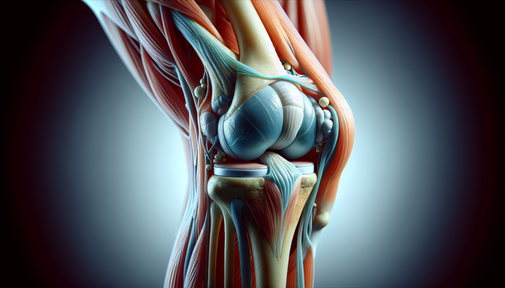
A Baker’s cyst, also known as a popliteal cyst, is a fluid-filled sac that forms in the popliteal fossa, which is located on the posterior aspect of the knee. It typically develops between the semimembranosus tendon and the medial head of the gastrocnemius muscle. The cyst is caused by an accumulation of synovial fluid that bulges into the back of the knee due to a valve-like effect between the joint space and the cyst.
Definition
A Baker’s cyst is defined as a synovial-lined fluid sac that arises from the gastrocnemio-semimembranosus bursa, resulting in a painful swelling behind the knee. The cyst forms when excess synovial fluid builds up and localizes in the popliteal fossa, often in association with an underlying knee condition such as osteoarthritis, rheumatoid arthritis, or a meniscal tear.
RELATED: Understanding Psoriasis: Symptoms, Causes, and Treatment Options
Anatomy
The popliteal fossa is a diamond-shaped space located at the back of the knee. It is bordered by the medial head of the gastrocnemius muscle and the semimembranosus tendon. In some cases, normal anatomic variants may include capsular openings or defects in the posterior knee joint capsule that allow for the formation of a Baker’s cyst. As the cyst enlarges, it can compress surrounding structures, leading to symptoms such as pain, tightness, and discomfort behind the knee.
Prevalence
Baker’s cysts are relatively common, with a prevalence ranging from 5% to 38% in the general population. The condition is most frequently encountered in adults between the ages of 35 and 70 years and is often associated with underlying knee pathologies. The prevalence of Baker’s cysts increases with age, likely due to the increased incidence of knee-bursal communication in older individuals. In children, Baker’s cysts tend to occur most frequently between the ages of 4 and 7 years, often arising as a primary condition rather than in association with an underlying knee disorder.
Causes and Risk Factors
A Baker’s cyst develops when excess synovial fluid accumulates in the bursa behind the knee. This accumulation of fluid can be caused by various factors, including knee joint issues, arthritis, injuries, and other medical conditions.
Several factors can contribute to the development of a Baker’s cyst:
- Knee joint issues: Any condition that causes inflammation or damage to the knee joint can lead to the formation of a Baker’s cyst. These conditions include meniscal tears, cartilage damage, and ligament injuries. The damaged tissues release excess synovial fluid, which can accumulate in the bursa and form a cyst.
- Arthritis: Inflammatory joint diseases such as rheumatoid arthritis and osteoarthritis can cause chronic inflammation in the knee joint. This inflammation leads to an increased production of synovial fluid, which can contribute to the development of a Baker’s cyst.
- Injuries: Trauma or injury to the knee, such as repetitive strain injuries, meniscus tears, hyperextensions, sprains, dislocations, and bone fractures, can cause swelling in the knee joint. The excess fluid produced during the healing process may drain into the bursa, leading to the formation of a Baker’s cyst.
- Other medical conditions: Certain medical conditions, such as gout and infectious arthritis, can cause inflammation in the knee joint, increasing the risk of developing a Baker’s cyst.
Risk factors for developing a Baker’s cyst include:
- Age: Baker’s cysts are more common in adults between the ages of 35 and 70 years.
- Arthritis: People with arthritis, particularly rheumatoid arthritis and osteoarthritis, are at a higher risk of developing Baker’s cysts.
- Knee injuries: Athletes and individuals who put significant pressure on their knees during work or hobbies are more susceptible to knee injuries, which can lead to the formation of Baker’s cysts.
Symptoms and Diagnosis
The most prominent symptom of a Baker’s cyst is a noticeable swelling or lump behind the knee. This swelling may be accompanied by pain, stiffness, and a reduced range of motion in the affected knee. In some cases, the cyst may cause no symptoms and may only be discovered during a physical examination or imaging study.
Common symptoms of a Baker’s cyst include:
- Swelling or a lump behind the knee
- Pain or tightness in the knee, especially when fully extending or flexing the joint
- Stiffness and reduced range of motion in the knee
- Swelling in the leg or calf (in case of cyst rupture)
It is essential to consult a healthcare provider if you experience any of these symptoms, as they can be similar to those of more serious conditions, such as a blood clot or tumor. Seeking prompt medical attention is crucial for an accurate diagnosis and appropriate treatment.
Diagnostic methods for Baker’s cysts typically involve a combination of physical examination and imaging studies. During the physical exam, the healthcare provider will assess the swelling behind the knee and evaluate the patient’s range of motion and symptoms. The provider may also check for signs of underlying conditions, such as arthritis or a knee injury.
RELATED: Eczema Breakdown: Types, Causes, Symptoms, and Treatments
To confirm the diagnosis and rule out other potential causes, imaging studies may be ordered, including:
- Ultrasound: This non-invasive test uses sound waves to create images of the soft tissues behind the knee, allowing the healthcare provider to visualize the cyst and assess its size and location.
- X-ray: While X-rays do not show the cyst itself, they can help identify any bony abnormalities or signs of arthritis that may be contributing to the development of the Baker’s cyst.
- MRI: Magnetic resonance imaging provides detailed images of the soft tissues and can help evaluate the extent of the cyst and any associated knee joint pathology.
By combining the findings from the physical examination and imaging studies, healthcare providers can accurately diagnose a Baker’s cyst and develop an appropriate treatment plan based on the underlying cause and severity of the condition.
Treatment Options and Management
Treatment for a Baker’s cyst depends on the severity of symptoms and the underlying cause. Conservative treatments are often the first line of defense, aiming to reduce pain and inflammation. These may include rest, ice application, compression, and elevation (RICE) of the affected knee. Over-the-counter pain medications such as ibuprofen or naproxen can help alleviate discomfort. Physical therapy exercises that focus on improving range of motion and strengthening the muscles around the knee may also be beneficial.
RELATED: Understanding Endometriosis: Symptoms and Causes Explained
If conservative measures fail to provide relief, medical interventions may be necessary. Corticosteroid injections into the knee joint can help reduce inflammation and pain. In some cases, the fluid from the cyst may be aspirated (drained) using a needle under ultrasound guidance. However, aspiration alone may not prevent the cyst from recurring if the underlying cause is not addressed.
Surgical options are typically reserved for cases where the Baker’s cyst causes severe symptoms or when conservative and medical treatments have been ineffective. Arthroscopic surgery can be performed to repair any damaged tissues within the knee joint, such as a torn meniscus or cartilage, which may be contributing to the formation of the cyst. In rare instances, open surgery may be necessary to remove a large or persistent cyst.
Conclusion
Baker’s cysts can have a significant impact on knee health and overall mobility. Understanding the causes, symptoms, and treatment options is crucial to manage this condition effectively. While these cysts often result from underlying knee issues or injuries, they can be addressed through a range of approaches, from simple rest and ice application to more advanced medical interventions. The key is to identify the root cause and tackle it head-on.
For those dealing with a Baker’s cyst, it’s essential to work closely with healthcare providers to develop a tailored treatment plan. This may involve lifestyle changes, physical therapy, or in some cases, surgical procedures to resolve the issue. By staying informed and proactive, individuals can take steps to alleviate discomfort and improve their quality of life. Remember, early detection and proper care are vital to prevent complications and ensure a speedy recovery.

