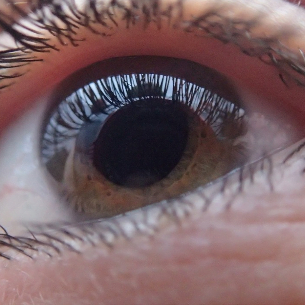
Glaucoma is a serious eye condition that affects millions of people worldwide. It’s often called the “silent thief of sight” because it can cause vision loss without noticeable symptoms in its early stages. This progressive disease damages the optic nerve, which is crucial for transmitting visual information from the eye to the brain. Understanding glaucoma is essential for early detection and effective management.
This article delves into the key aspects of glaucoma to help readers grasp its importance. It covers the different types and causes of glaucoma, how to spot its symptoms, and the methods used to diagnose it. The piece also explores various treatment options available and offers insights on living with the condition. By providing this comprehensive overview, we aim to raise awareness and encourage regular eye check-ups to catch glaucoma early.
Understanding Glaucoma: Types and Causes


Glaucoma is a group of eye diseases that damage the optic nerve, which is crucial for transmitting visual information from the eye to the brain. The two main types of glaucoma are open-angle glaucoma and angle-closure glaucoma. Each type has different characteristics and causes.
Open-angle glaucoma
Open-angle glaucoma is the most common form in the United States, accounting for about 90% of all glaucoma cases. In this type, the drainage angle between the iris and cornea remains open, but the fluid cannot drain properly, causing a gradual increase in eye pressure. Symptoms may not be noticeable until significant vision loss has occurred.
Primary open-angle glaucoma is often hereditary, and risk factors include age, family history, and being of African, Asian, or Hispanic descent. Secondary open-angle glaucoma can be caused by other medical conditions, medications, or eye injuries.
Angle-closure glaucoma
RELATED: Everything About Folliculitis: From Causes to Treatment
Angle-closure glaucoma, also known as narrow-angle or acute glaucoma, occurs when the outer edge of the iris blocks the drainage angle, causing a rapid buildup of fluid and a sudden increase in eye pressure. This type of glaucoma is a medical emergency and can cause blindness within a few days if left untreated.
Risk factors for angle-closure glaucoma include being of Asian descent, having a family history of the condition, and having certain eye anatomy, such as a shallow anterior chamber or a thick lens.
Normal-tension glaucoma
Normal-tension glaucoma, a type of open-angle glaucoma, occurs when there is damage to the optic nerve despite having normal eye pressure. The exact cause is unknown, but it may be related to reduced blood flow to the optic nerve, structural weakness of the lamina cribrosa, or low blood pressure.
Risk factors for normal-tension glaucoma include being of Japanese ancestry, having a family history of the condition, and having certain heart problems or low blood pressure.
Risk factors
In addition to the risk factors mentioned for each type of glaucoma, other factors that can increase the risk of developing the disease include:
- Age over 60 years old
- Thin central corneas
- Extreme nearsightedness or farsightedness
- Prolonged use of corticosteroid medications
- Systemic conditions such as diabetes, hypertension, and heart disease
Understanding the different types of glaucoma and their associated risk factors is essential for early detection and effective management. Regular comprehensive eye exams can help identify glaucoma in its early stages, allowing for timely treatment to prevent vision loss.
Recognizing Glaucoma Symptoms
Glaucoma is often called the “silent thief of sight” because it can cause significant damage to the optic nerve without noticeable symptoms in its early stages. However, being aware of potential warning signs and symptoms can help with early detection and timely treatment.
Early warning signs
In the early stages of glaucoma, especially open-angle glaucoma, there may be no apparent symptoms. This lack of symptoms makes regular comprehensive eye exams crucial for early detection. As the disease progresses, some signs and symptoms may include:
- Gradual loss of peripheral (side) vision
- Patchy blind spots in your central or peripheral vision
- Tunnel vision in advanced stages
Symptoms of acute angle-closure glaucoma
Acute angle-closure glaucoma is a medical emergency that requires immediate attention. Symptoms may include:
- Severe eye pain
- Nausea and vomiting
- Sudden onset of visual disturbance, often in low light
- Blurred vision
- Halos around lights
- Redness in the eye
When to seek medical attention
If you experience any symptoms of acute angle-closure glaucoma, seek immediate medical care. This condition can cause permanent vision loss within a day if left untreated.
For other types of glaucoma, it is essential to have regular eye exams to monitor eye health and detect any changes early on. The frequency of these exams may vary based on age, family history, and other risk factors. Your eye care professional can recommend an appropriate screening schedule for you.
Remember, early detection and treatment are key to preventing vision loss from glaucoma. If you notice any changes in your vision or experience any concerning symptoms, do not hesitate to consult with your eye care provider.
Diagnosing Glaucoma
Diagnosing glaucoma involves a comprehensive eye examination that includes several tests to evaluate the health of the optic nerve and measure the intraocular pressure (IOP). Early detection is crucial for preventing vision loss, as glaucoma often progresses without noticeable symptoms in its early stages.
RELATED: Flea Bites in Humans: Symptoms, Treatment, and Prevention
Comprehensive eye examination
A comprehensive eye examination is the first step in diagnosing glaucoma. During this exam, the eye doctor will:
- Review the patient’s medical history and any risk factors for glaucoma
- Assess visual acuity using an eye chart
- Examine the anterior segment of the eye using a slit lamp
- Evaluate the optic nerve for signs of damage
- Measure the intraocular pressure (IOP) using tonometry
Tonometry
Tonometry is a test that measures the pressure inside the eye. There are several types of tonometry, including:
- Applanation tonometry: This is the most common and accurate method. It involves gently touching the surface of the eye with a small probe after numbing the eye with drops.
- Non-contact tonometry (air puff test): This method uses a rapid air pulse to flatten the cornea and measure the IOP without touching the eye.
Elevated IOP is a significant risk factor for glaucoma, although not all individuals with high IOP develop the disease.
Visual field test
A visual field test, also known as perimetry, assesses peripheral vision and can detect areas of vision loss caused by glaucoma. During the test, the patient focuses on a central point while lights of varying intensities appear in their peripheral vision. The patient responds when they see a light, and the results are recorded on a chart.
There are two main types of visual field tests:
- Automated static perimetry: This is the most common type, where the patient sits in front of a machine and presses a button when they see a light.
- Kinetic perimetry (Goldmann perimetry): In this test, the examiner moves a light target from the periphery towards the center of vision, and the patient indicates when they first see the light.
Repeating visual field tests over time can help monitor the progression of glaucoma.
Optical coherence tomography (OCT)
Optical coherence tomography (OCT) is a non-invasive imaging technology that provides high-resolution, cross-sectional images of the retina and optic nerve. OCT can measure the thickness of the retinal nerve fiber layer (RNFL) and the ganglion cell complex (GCC), which are often damaged in glaucoma.
OCT is useful for:
- Detecting early signs of glaucoma before visual field defects develop
- Monitoring the progression of glaucoma over time
- Helping to differentiate between glaucoma and other optic neuropathies
Advancements in OCT technology, such as spectral-domain OCT (SD-OCT) and swept-source OCT (SS-OCT), have improved the resolution and speed of scans, allowing for more detailed assessment of the optic nerve and retina.
In summary, diagnosing glaucoma requires a combination of tests performed during a comprehensive eye examination. Tonometry, visual field testing, and optical coherence tomography are essential tools for detecting and monitoring glaucoma, enabling early intervention to preserve vision.
Treatment Options for Glaucoma
The goal of glaucoma treatment is to lower intraocular pressure (IOP) and prevent further damage to the optic nerve. There are several treatment options available, including medications, laser therapy, surgery, and lifestyle changes. The choice of treatment depends on the type and severity of glaucoma, as well as individual patient factors.
Medications
Prescription eye drops are the most common initial treatment for glaucoma. These medications work by either decreasing the production of aqueous humor or increasing its outflow from the eye. Some examples of glaucoma eye drops include:
- Prostaglandin analogs
- Rho kinase inhibitors (e.g., netarsudil)
- Nitric oxides
- Miotic or cholinergic agents
- Alpha-adrenergic agonists
- Beta blockers
- Carbonic anhydrase inhibitors (e.g., dorzolamide)
In some cases, oral medications such as carbonic anhydrase inhibitors may be prescribed if eye drops alone are not effective in lowering IOP.
Laser therapy
Laser treatments can be used as a first-line therapy or in combination with medications to lower IOP. Some common laser procedures for glaucoma include:
- Argon laser trabeculoplasty (ALT)
- Selective laser trabeculoplasty (SLT)
- Laser peripheral iridotomy (LPI)
- Cyclophotocoagulation
These procedures work by either improving the outflow of aqueous humor or reducing its production. Laser treatments are usually performed in an outpatient setting and have a relatively short recovery time.
Surgery
If medications and laser therapy fail to control IOP, surgical interventions may be necessary. The most common surgical procedures for glaucoma are:
- Trabeculectomy: This procedure involves creating a small flap in the sclera to allow aqueous humor to drain from the eye, thereby lowering IOP.
- Glaucoma drainage devices (e.g., Ahmed, Baerveldt, Molteno implants): These devices are inserted into the eye to create an alternate pathway for aqueous humor drainage.
- Minimally invasive glaucoma surgery (MIGS): These procedures, such as iStent, Trabectome, and XEN Gel Stent, are less invasive than traditional surgeries and aim to improve aqueous humor outflow with fewer complications.
Surgical treatments for glaucoma are typically performed in an operating room setting and may require a longer recovery period compared to laser therapy.
Lifestyle changes
In addition to medical and surgical interventions, certain lifestyle modifications may help manage glaucoma and promote overall eye health:
- Regular exercise: Moderate physical activity has been shown to lower IOP and improve blood flow to the optic nerve.
- Healthy diet: Eating a diet rich in fruits, vegetables, and omega-3 fatty acids may help protect against glaucoma progression.
- Avoiding head-down positions: Certain yoga poses and sleeping with the head elevated may help reduce IOP spikes.
- Protecting the eyes from trauma: Wearing protective eyewear during sports and other high-risk activities can help prevent eye injuries that may worsen glaucoma.
It is essential for patients with glaucoma to adhere to their prescribed treatment plan and attend regular follow-up appointments with their eye care provider to monitor disease progression and adjust therapy as needed.
Living with Glaucoma: Coping Strategies
Living with glaucoma can be challenging, but adopting effective coping strategies can help manage the condition and maintain a good quality of life. Regular check-ups, medication adherence, support groups, and adapting your environment are key aspects of successfully coping with glaucoma.
Regular check-ups with your eye care professional are crucial for monitoring the progression of glaucoma and adjusting treatment plans as needed. During these appointments, your doctor will assess your intraocular pressure, examine the optic nerve, and perform visual field tests to detect any changes in your vision. Consistent follow-up ensures that any potential issues are identified and addressed promptly, helping to preserve your sight.
Medication adherence is another critical component of managing glaucoma. Consistently taking prescribed eye drops or oral medications as directed by your doctor helps to control intraocular pressure and slow the progression of the disease. Set reminders, use pill organizers, or enlist the help of family members to ensure that you never miss a dose. If you experience side effects or have difficulty administering the drops, discuss these concerns with your doctor to find a solution that works for you.
Joining a support group can provide valuable emotional support and practical advice for coping with glaucoma. Connecting with others who share similar experiences can help you feel less isolated and more empowered to manage your condition. Support groups offer a platform to exchange information, share coping strategies, and learn about the latest research and treatment options. Many organizations, such as the Glaucoma Research Foundation and the American Glaucoma Society, offer online and in-person support groups.
RELATED: Comprehensive Overview of Depression: Causes, Symptoms, and Treatments
Adapting your environment can make daily tasks easier and safer for those living with glaucoma. Some simple modifications include:
- Improving lighting: Ensure that your home and workspace have adequate lighting to reduce eye strain and improve visibility.
- Reducing glare: Use window coverings or anti-glare filters on electronic devices to minimize glare, which can be particularly bothersome for those with glaucoma.
- Organizing your space: Keep frequently used items in easily accessible locations and maintain a clutter-free environment to reduce the risk of accidents.
- Using assistive devices: Magnifying glasses, large-print books, and talking watches can help you maintain independence and engage in activities you enjoy.
By incorporating these coping strategies into your daily life, you can effectively manage glaucoma and maintain a high quality of life. Remember, your eye care team is there to support you every step of the way, so don’t hesitate to reach out for guidance and assistance whenever needed.
Conclusion
Glaucoma poses significant challenges to eye health, but understanding its complexities can lead to better management and outcomes. This article has shed light on the various types of glaucoma, their symptoms, diagnostic methods, and treatment options. By raising awareness about this condition, we hope to encourage regular eye check-ups and prompt medical attention when needed. Early detection and proper care are key to preserving vision and maintaining a good quality of life for those affected by glaucoma.
Living with glaucoma requires a multi-faceted approach, combining medical treatments with lifestyle adjustments. By following prescribed treatments, attending regular check-ups, and making necessary adaptations in daily life, individuals with glaucoma can effectively manage their condition. Remember, you’re not alone in this journey – support groups and healthcare professionals are there to help. With the right knowledge and support, people with glaucoma can continue to lead fulfilling lives while taking care of their eye health.

