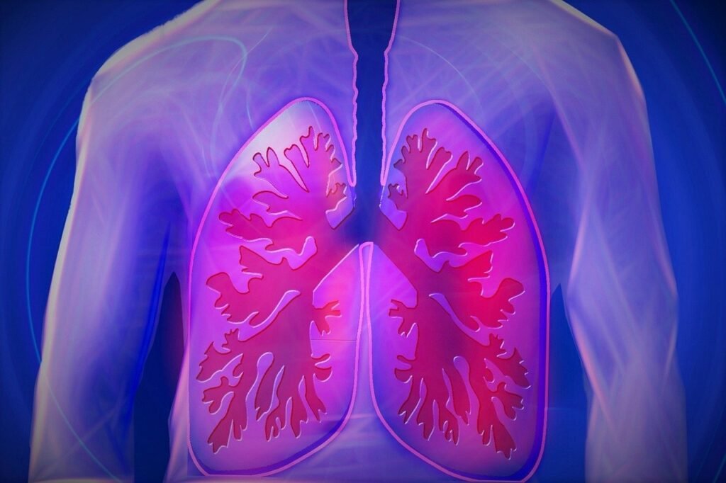
A pneumothorax, commonly known as a collapsed lung, is a serious medical condition that requires immediate attention. This occurs when air leaks into the space between the lung and chest wall, causing the lung to collapse partially or completely. Understanding the causes, symptoms, and treatment options for pneumothorax is crucial for both medical professionals and the general public.
Pneumothorax can happen spontaneously or result from chest injuries, medical procedures, or underlying lung diseases. This article aims to shed light on how to recognize the signs of a collapsed lung and the various approaches to treat it. It will cover the different types of pneumothorax, how doctors diagnose the condition, and the recovery process patients can expect. By exploring these aspects, readers will gain valuable insights into this potentially life-threatening condition and the importance of seeking prompt medical care.
Types and Causes of Pneumothorax
Pneumothorax can be classified into three main types based on their underlying causes: spontaneous pneumothorax, traumatic pneumothorax, and iatrogenic pneumothorax. Each type has its own set of risk factors and characteristics.
Spontaneous Pneumothorax
Spontaneous pneumothorax occurs without an obvious etiology and can be further divided into primary and secondary types. Primary spontaneous pneumothorax happens in individuals with no known history of lung disease, often due to the rupture of abnormal air pockets in the lung called blebs. Risk factors for primary spontaneous pneumothorax include:
- Tall, thin body habitus, especially in males
- Smoking
- Family history of pneumothorax
- Connective tissue disorders like Marfan syndrome
Secondary spontaneous pneumothorax is associated with pre-existing lung diseases such as:
- Chronic obstructive pulmonary disease (COPD)
- Asthma
- Cystic fibrosis
- Pneumonia
- Lung cancer
- Interstitial lung diseases (e.g., idiopathic pulmonary fibrosis, sarcoidosis)
RELATED: Melasma Management: Causes, Symptoms, and Best Treatments
Traumatic Pneumothorax
Traumatic pneumothorax results from chest injuries, which can be either penetrating or non-penetrating. Penetrating trauma, such as stab wounds or gunshot wounds, can directly injure the lung and allow air to enter the pleural space. Non-penetrating or blunt trauma can cause rib fractures or lung lacerations, leading to air leakage into the pleural space.
Iatrogenic Pneumothorax
Iatrogenic pneumothorax occurs as a complication of medical procedures. Some common causes include:
- Transthoracic needle aspiration or biopsy
- Central venous catheterization (e.g., subclavian or jugular vein)
- Thoracentesis
- Mechanical ventilation
- Cardiopulmonary resuscitation
The risk of iatrogenic pneumothorax varies depending on the procedure and the patient’s underlying condition. For example, patients with COPD or those requiring mechanical ventilation are at a higher risk of developing pneumothorax.
Understanding the types and causes of pneumothorax is crucial for prompt recognition, appropriate management, and prevention of this potentially life-threatening condition.
Identifying Pneumothorax Symptoms
The symptoms of a pneumothorax can vary depending on the severity and underlying cause. In some cases, a collapsed lung may not cause any noticeable symptoms, while in others, it can lead to life-threatening complications. Recognizing the signs and symptoms of a pneumothorax is crucial for prompt medical attention and treatment.
The most common symptoms of a pneumothorax include sudden chest pain, often described as sharp or stabbing, that worsens with breathing or coughing. Shortness of breath, rapid breathing, and a rapid heart rate may also occur as the body tries to compensate for the reduced oxygen supply. In severe cases, the skin may take on a bluish tint (cyanosis) due to a lack of oxygen.
Other symptoms may include fatigue, a dry cough, and a feeling of tightness in the chest. In some cases, individuals may experience a sensation of air bubbling under the skin around the chest or neck, known as subcutaneous emphysema.
RELATED: Mastitis: Detailed Insights into Causes, Symptoms, and Treatments
It is essential to seek immediate medical attention if you suspect a pneumothorax, as it can quickly progress to a life-threatening condition. Complications of a collapsed lung may include:
- Tension pneumothorax: A severe form of pneumothorax where air continues to accumulate in the pleural space, putting pressure on the heart and other organs.
- Hypoxemia: Low levels of oxygen in the blood, which can lead to organ damage if left untreated.
- Respiratory failure: A condition where the lungs cannot provide sufficient oxygen to the body or remove carbon dioxide effectively.
By familiarizing yourself with the symptoms of a pneumothorax and seeking prompt medical care, you can reduce the risk of serious complications and ensure a better outcome.
Medical Evaluation and Diagnosis
Prompt diagnosis of a pneumothorax is crucial for timely treatment and prevention of complications. The evaluation process typically involves a combination of patient history, physical examination, and diagnostic imaging.
Patient History
A thorough patient history is essential in diagnosing pneumothorax. Key information to gather includes:
- Onset and duration of symptoms such as chest pain, shortness of breath, and cough
- Presence of underlying lung diseases like COPD, asthma, or cystic fibrosis
- Recent medical procedures, including lung biopsies or central line placements
- History of trauma to the chest
- Smoking habits and exposure to environmental smoke
Physical Examination
Physical examination findings that suggest pneumothorax include:
- Unequal breath sounds, with decreased or absent breath sounds on the affected side
- Hyperresonance to percussion over the affected hemithorax
- Decreased chest wall movement on the affected side
- Tachycardia, tachypnea, hypoxia, and hypotension in severe cases
Diagnostic Imaging
Imaging studies are crucial for confirming the diagnosis and assessing the extent of pneumothorax. The most commonly used modalities are:
- Chest X-ray: An upright posteroanterior chest radiograph is the initial imaging study of choice. It can demonstrate the presence of air in the pleural space and the degree of lung collapse. In supine patients, the deep sulcus sign may be the only indicator of pneumothorax.
- Computed Tomography (CT): CT scans provide more detailed images and can detect small pneumothoraces not visible on chest X-rays. They are particularly useful in evaluating patients with underlying lung diseases or traumatic injuries.
- Ultrasound: Bedside ultrasound is increasingly used as a rapid, non-invasive tool for diagnosing pneumothorax, especially in critical care settings. The absence of lung sliding and the presence of a lung point are highly suggestive of pneumothorax.
By integrating information from the patient history, physical examination, and diagnostic imaging, healthcare providers can accurately diagnose pneumothorax and initiate appropriate treatment. Early recognition and intervention are key to preventing the progression of pneumothorax and minimizing the risk of life-threatening complications such as tension pneumothorax.
Treatment Approaches and Recovery
The treatment of pneumothorax depends on its severity and underlying cause. In general, the goals are to remove air from the pleural space, allow the lung to re-expand, and prevent recurrence. Treatment options range from observation to surgical intervention.
Emergency Care
In the prehospital setting, tension pneumothorax requires immediate needle decompression to prevent cardiac arrest. This involves inserting a large-bore needle into the second intercostal space at the midclavicular line to allow trapped air to escape. A one-way valve should be attached to prevent air reentry.
Upon arrival at the hospital, a chest tube is inserted to continuously drain air and allow the lung to re-expand. Supplemental oxygen is administered to promote the absorption of pleural air. Patients with large or symptomatic pneumothoraces typically require chest tube placement, while those with small, stable pneumothoraces may be managed with observation and supplemental oxygen.
Long-term Management
After initial treatment, long-term management focuses on preventing recurrence. Strategies include:
- Pleurodesis: Chemically or surgically irritating the pleural space to promote adhesion and prevent air accumulation.
- Video-assisted thoracoscopic surgery (VATS): Minimally invasive procedure to resect blebs, perform pleurodesis, and close air leaks.
- Open thoracotomy: More invasive surgery for complex cases or failed VATS.
The choice of intervention depends on factors such as the patient’s overall health, underlying lung disease, and pneumothorax recurrence risk.
RELATED: The Complete Guide to Macular Degeneration: Symptoms and Diagnosis
Prevention Strategies
Preventing pneumothorax recurrence is crucial, particularly in high-risk populations. Strategies include:
- Smoking cessation: Reduces the risk of spontaneous pneumothorax and improves overall lung health.
- Avoiding high-risk activities: Scuba diving, flying, and contact sports may be restricted in patients with a history of pneumothorax.
- Prompt treatment of underlying lung diseases: Optimal management of conditions like COPD and asthma can reduce the risk of secondary pneumothorax.
- Pleurodesis or surgical intervention: Considered for patients with recurrent pneumothoraces or high-risk occupations.
Close follow-up with a pulmonologist is essential to monitor for recurrence and manage underlying lung conditions. With appropriate treatment and prevention strategies, most patients with pneumothorax can achieve a full recovery and minimize the risk of future episodes.
Conclusion
Recognizing and treating pneumothorax quickly is key to preventing serious complications. This article has shed light on the various types of collapsed lungs, their causes, and the telltale signs to watch out for. By understanding the symptoms and seeking prompt medical care, patients can improve their chances of a full recovery. The diagnostic process, involving patient history, physical exams, and imaging studies, plays a crucial role in pinpointing the condition and guiding treatment decisions.
Managing pneumothorax involves a range of approaches, from simple observation to more invasive procedures like chest tube insertion or surgery. The choice of treatment depends on the severity and underlying cause of the condition. To wrap up, preventing recurrence is just as important as the initial treatment. Strategies such as quitting smoking, avoiding high-risk activities, and keeping underlying lung conditions in check can go a long way in reducing the risk of future episodes. With the right care and preventive measures, most people with pneumothorax can look forward to a healthy future.

