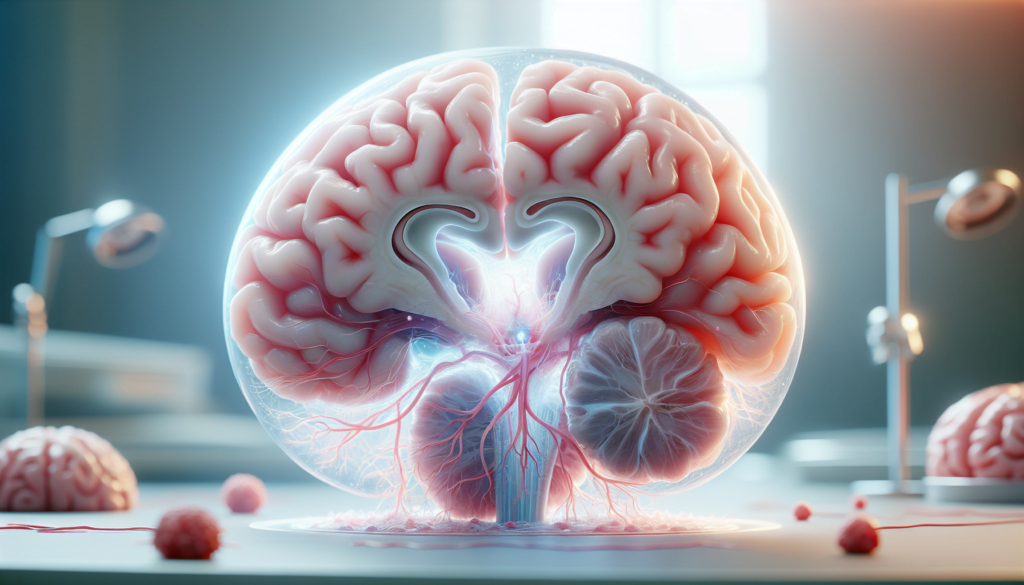
Hydrocephalus is a complex neurological condition that affects thousands of people worldwide. This disorder, characterized by an abnormal buildup of cerebrospinal fluid in the brain, can have a significant impact on an individual’s quality of life. From infants to adults, hydrocephalus can cause a range of symptoms and complications, making it a crucial area of study in neurology and neurosurgery.
Understanding hydrocephalus is essential to improve diagnosis, treatment, and long-term management of this condition. This article aims to provide a comprehensive overview of hydrocephalus, exploring its causes, symptoms, and available treatment options. By delving into the latest research and medical advancements, we hope to shed light on this often misunderstood condition and offer valuable insights to those affected by it or interested in learning more about this challenging neurological disorder.
Understanding Hydrocephalus


Hydrocephalus is a complex neurological disorder characterized by an abnormal accumulation of cerebrospinal fluid (CSF) within the ventricles of the brain. This buildup of fluid causes the ventricles to enlarge, putting pressure on the surrounding brain tissue and potentially leading to a range of symptoms and complications.
What is Hydrocephalus?
The term “hydrocephalus” is derived from the Greek words “hydro,” meaning water, and “cephalus,” meaning head. Despite this literal translation, the condition actually involves an excess of CSF rather than water. CSF is a clear, colorless fluid that surrounds and cushions the brain and spinal cord, providing essential protection and nourishment.
In a healthy brain, CSF is produced, circulated, and absorbed at a balanced rate. However, in hydrocephalus, this balance is disrupted, leading to an accumulation of fluid within the ventricles. This disruption can occur due to an obstruction in the flow of CSF, problems with absorption, or, in rare cases, an overproduction of the fluid.
Types of Hydrocephalus
- Communicating hydrocephalus: This type occurs when the flow of CSF is blocked after it exits the ventricles, while the passages between the ventricles remain open.
- Non-communicating (obstructive) hydrocephalus: In this type, the flow of CSF is blocked along one or more of the narrow passages connecting the ventricles.
- Normal pressure hydrocephalus (NPH): This form of communicating hydrocephalus often affects older adults and is characterized by the gradual onset of symptoms such as difficulty walking, cognitive impairment, and urinary incontinence.
- Hydrocephalus ex-vacuo: This condition results from brain damage caused by stroke or injury, leading to the shrinkage of brain tissue and the subsequent enlargement of the ventricles.
RELATED: Anal Fissures: Effective Treatments and Healing Tips
The Role of Cerebrospinal Fluid
CSF plays a crucial role in maintaining proper brain function by:
- Acting as a shock absorber, protecting the brain and spinal cord from injury
- Delivering nutrients to the brain and removing waste products
- Regulating intracranial pressure by flowing between the cranium and spine
When the delicate balance of CSF production, circulation, and absorption is disrupted, as in the case of hydrocephalus, it can have a significant impact on brain function and overall health. Understanding the underlying mechanisms of hydrocephalus is essential for developing effective diagnostic and treatment strategies to manage this challenging condition.
Causes and Risk Factors
Hydrocephalus can be caused by a variety of factors, including congenital abnormalities, acquired conditions, and certain risk factors that increase the likelihood of developing the disorder.
Congenital Causes
Congenital hydrocephalus develops before or shortly after birth due to abnormalities in brain development. Common causes include:
- Blockage of the cerebral aqueduct, the narrow passage between the third and fourth ventricles of the brain
- Neural tube defects such as spina bifida
- Dandy-Walker syndrome, characterized by an enlarged fourth ventricle
- Genetic abnormalities that disrupt the flow of cerebrospinal fluid (CSF)
Maternal infections during pregnancy, such as rubella and syphilis, can also lead to congenital hydrocephalus by causing inflammation in the fetal brain tissue.
Acquired Causes
Acquired hydrocephalus develops after birth as a result of various neurological conditions, including:
- Head trauma or brain injury
- Brain tumors that obstruct CSF flow
- Intraventricular hemorrhage, common in premature infants
- Central nervous system infections like bacterial meningitis or mumps
These conditions can cause scarring, inflammation, or blockages within the ventricular system, disrupting the normal circulation and absorption of CSF.
Risk Factors
Several factors can increase the risk of developing hydrocephalus:
- Premature birth and its associated complications
- Family history of hydrocephalus
- Congenital heart disease
- Spinal cord abnormalities
- Certain genetic disorders, such as X-linked hydrocephalus
Understanding the causes and risk factors of hydrocephalus is crucial for early detection, prevention, and appropriate management of this complex neurological disorder.
Symptoms and Diagnosis
The symptoms of hydrocephalus can vary depending on the age of onset. In infants, the most noticeable sign is a rapid increase in head circumference or an unusually large head size. Other symptoms may include:
- Bulging or tense soft spot on the top of the head (fontanel)
- Downward-gazing eyes (sunsetting of the eyes)
- Irritability and high-pitched crying
- Poor feeding and vomiting
- Seizures and sleepiness
In older children and adults, symptoms may be less obvious and can include:
- Headaches, often accompanied by nausea and vomiting
- Blurred or double vision
- Balance and coordination problems
- Urinary incontinence or frequent urination
- Lethargy and drowsiness
- Personality changes and decline in school or work performance
- Memory loss and difficulty concentrating
To diagnose hydrocephalus, healthcare professionals conduct a thorough neurological examination and may recommend neuropsychological testing to assess cognitive function. Brain imaging tests play a crucial role in confirming the diagnosis and determining the underlying cause of the condition.
RELATED: Avoidant/Restrictive Food Intake Disorder (ARFID): Symptoms and Treatment
Diagnostic Methods
- Ultrasound: This non-invasive technique uses sound waves to create images of the brain’s ventricles. It is often the first test performed on infants because their fontanels allow for easy access. Ultrasound can also detect hydrocephalus before birth during prenatal examinations.
- Magnetic Resonance Imaging (MRI): MRI scans provide detailed images of the brain, allowing doctors to evaluate the size of the ventricles, assess the flow of cerebrospinal fluid, and identify any underlying conditions such as tumors or congenital malformations. Sedation may be necessary for young children to ensure they remain still during the scan.
- Computed Tomography (CT) Scan: CT scans use X-rays to create cross-sectional images of the brain. They can quickly reveal enlarged ventricles and are often used in emergency situations. However, CT scans expose the patient to a small amount of radiation and provide less detailed images compared to MRI scans.
By combining the results of neurological examinations, neuropsychological testing, and brain imaging, healthcare professionals can accurately diagnose hydrocephalus and develop an appropriate treatment plan tailored to the individual’s needs.
Treatment Options and Outlook
The primary treatment for hydrocephalus involves surgical interventions to relieve the pressure on the brain caused by the excess cerebrospinal fluid (CSF). The two main surgical options are shunt systems and endoscopic third ventriculostomy (ETV).
Surgical Interventions
Shunt systems are the most common treatment for hydrocephalus. A shunt is a medical device that consists of a long, flexible tube with a valve that drains the excess CSF from the brain to another part of the body, such as the abdomen or heart, where it can be absorbed naturally. Shunts are usually placed in the brain’s ventricles and require lifelong monitoring and potential revisions.
Shunt Systems
There are different types of shunt systems, including:
- Ventriculoperitoneal (VP) shunts
- Ventriculoatrial (VA) shunts
- Ventriculopleural (VPL) shunts
- Lumboperitoneal (LP) shunts
Shunts can have fixed or adjustable (programmable) valves, which regulate the pressure and flow of CSF. Complications associated with shunts include malfunction, infection, and over- or under-drainage.
Endoscopic Third Ventriculostomy
ETV is an alternative surgical procedure that involves creating a small hole in the floor of the third ventricle, allowing the CSF to bypass any obstruction and flow directly into the brain’s subarachnoid space. ETV is suitable for certain types of hydrocephalus and has a lower risk of infection compared to shunts.
RELATED: What Are Autoimmune Diseases? Types, Symptoms, and Treatments
Long-term Management
Individuals with hydrocephalus require long-term management and follow-up care to monitor their condition and address any complications. This may include regular imaging, neurological exams, and shunt function assessments. Rehabilitation therapies, such as physical therapy and occupational therapy, may be recommended to help improve symptoms and quality of life.
Prognosis
The prognosis for individuals with hydrocephalus depends on various factors, including the underlying cause, timeliness of diagnosis and treatment, and the presence of associated disorders. With appropriate treatment and management, many people with hydrocephalus can lead normal lives with few limitations. However, some may experience ongoing challenges related to cognitive development, learning, and physical abilities. Early diagnosis and intervention are crucial for optimal outcomes and minimizing the risk of long-term complications.
Conclusion
Hydrocephalus has a significant impact on individuals and families, presenting unique challenges in diagnosis, treatment, and long-term care. The complexity of this condition, with its various causes and manifestations, underscores the need for ongoing research and medical advancements. Early detection and timely intervention are key to improving outcomes and quality of life for those affected by this neurological disorder.
While surgical treatments like shunts and endoscopic procedures offer hope, they also come with potential complications and require lifelong monitoring. The journey of living with hydrocephalus often involves a multidisciplinary approach, combining medical care with rehabilitation therapies. As our understanding of this condition grows, so does the potential to develop more effective treatments and support strategies, giving hope to those navigating the challenges of hydrocephalus.

