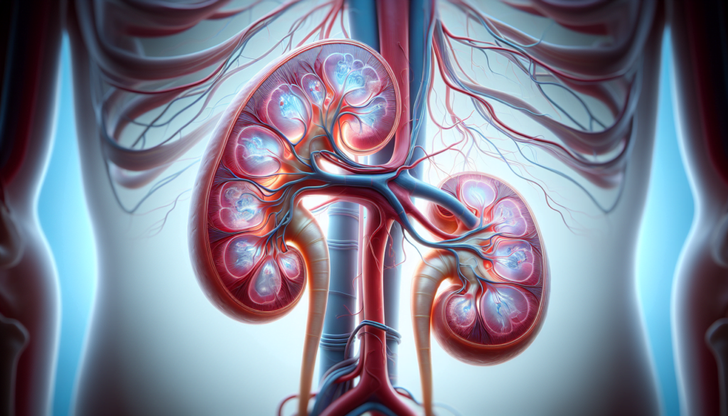
Hydronephrosis is a condition that affects countless individuals worldwide, causing distress and discomfort. This medical issue occurs when urine builds up in the kidneys, leading to swelling and potential damage. Understanding hydronephrosis is crucial for early detection and proper management, as it can have a significant impact on kidney function and overall health.
In this article, we’ll explore the key aspects of hydronephrosis, including its common causes and telltale symptoms. We’ll also delve into the diagnostic methods used by healthcare professionals to identify this condition. Additionally, we’ll discuss effective treatments and management strategies to help patients cope with hydronephrosis and prevent complications. By the end, readers will have a comprehensive understanding of this important urological issue.
Understanding Hydronephrosis
Hydronephrosis is a condition that occurs when urine accumulates in the kidneys, causing them to swell and become distended. This happens due to an obstruction of the outflow of urine distal to the renal pelvis. The condition can affect one or both kidneys and can be acute or chronic, unilateral or bilateral, and physiologic or pathologic.
Definition and Prevalence
Hydronephrosis is defined as the distention of the renal calyces and pelvis with urine as a result of obstruction of the outflow of urine distal to the renal pelvis. It is a common clinical condition encountered not only by urologists but also by emergency medicine specialists and primary care physicians. The presence of hydronephrosis can be physiologic or pathologic, and it may be acute or chronic, unilateral or bilateral.
Hydronephrosis is a normal finding in pregnant women, with the renal pelvises and caliceal systems dilated as a result of progesterone effects and mechanical compression of the ureters at the pelvic brim. This dilatation is more prominent on the right side than the left side and is seen in up to 80% of pregnant women.
RELATED: What Are Autoimmune Diseases? Types, Symptoms, and Treatments
Anatomy of the Urinary System
The urinary system is a complex, multi-component organ system that has the primary function of maintaining body homeostasis by regulating the volume of body fluid, electrolyte balance, and excretion of metabolic end products through urine. Anatomically, it includes the kidneys, ureters, urinary bladder, and urethra. Each kidney has an outer cortex and an inner medulla, which is formed into renal pyramids that extend into the renal pelvis, which is continued as the ureter.
Types of Hydronephrosis
Hydronephrosis can be classified as obstructive or non-obstructive. Obstructive hydronephrosis is caused by a blockage in the urinary tract, such as a kidney stone, tumor, or stricture. Non-obstructive hydronephrosis, on the other hand, is not caused by a blockage but rather by other factors such as vesicoureteral reflux or a neurogenic bladder.
Hydronephrosis can also be classified as unilateral or bilateral. Unilateral hydronephrosis affects only one kidney, while bilateral hydronephrosis affects both kidneys. Bilateral hydronephrosis is usually caused by an obstruction distal to the urinary bladder, with posterior urethral valves being the most common cause.
Common Causes of Hydronephrosis
Hydronephrosis can result from a variety of factors that obstruct or impede the flow of urine from the kidney to the bladder. These causes can be classified into two main categories: obstructive and non-obstructive.
Obstructive Causes
Obstructive hydronephrosis occurs when there is a physical blockage in the urinary tract that prevents urine from flowing freely. Some common obstructive causes include:
- Kidney stones: Stones can form in the kidney and become lodged in the ureter, blocking the flow of urine.
- Tumors: Cancerous or benign growths in the kidney, ureter, bladder, or prostate can compress or obstruct the urinary tract.
- Ureteropelvic junction (UPJ) obstruction: A narrowing or kinking of the ureter where it joins the kidney can impede urine flow.
- Posterior urethral valves: This congenital condition, occurring only in boys, involves a narrowing of the urethra that prevents free urine flow from the bladder.
Non-Obstructive Causes
Non-obstructive hydronephrosis is not caused by a physical blockage but rather by other factors that affect urine flow. These include:
- Vesicoureteral reflux: An abnormal backflow of urine from the bladder into the ureter and up to the kidney, often due to an abnormality in how the ureter connects with the bladder.
- Neurogenic bladder: Nerve problems that disrupt the normal functioning of the urinary system.
Risk Factors
Certain factors can increase the risk of developing hydronephrosis:
- Pregnancy: The growing uterus can compress the ureters, leading to a temporary form of hydronephrosis.
- Urinary tract infections: Recurrent infections can cause inflammation and scarring, potentially leading to obstruction.
- Congenital abnormalities: Some individuals are born with structural abnormalities of the urinary tract that predispose them to hydronephrosis.
Understanding the underlying cause of hydronephrosis is crucial for determining the most appropriate treatment approach and preventing potential complications.
Key Symptoms and Diagnosis
The symptoms of hydronephrosis can vary depending on the underlying cause and severity of the condition. In some cases, individuals may not experience any noticeable symptoms, especially if the hydronephrosis is mild or in its early stages.
Common Symptoms
When symptoms do occur, they may include:
- Pain in the lower abdomen, back, or flank area
- Nausea and vomiting
- Frequent urination or difficulty urinating
- Weak urine stream
- Urinary tract infections
- Fever and chills (in cases of infection)
Infants with hydronephrosis may exhibit symptoms such as lack of appetite and frequent urinary tract infections.
Diagnostic Procedures
To diagnose hydronephrosis, healthcare professionals rely on various imaging techniques and tests:
- Ultrasound: This non-invasive imaging method uses sound waves to create pictures of the kidneys and urinary tract. It is often the first test performed to detect hydronephrosis, especially in pregnant women and infants.
- CT scan: A computed tomography (CT) scan provides detailed cross-sectional images of the kidneys and surrounding structures. It is highly sensitive and specific in identifying the cause of hydronephrosis, such as kidney stones or tumors.
- Intravenous pyelogram (IVP): This test involves injecting a contrast dye into the bloodstream, which then travels through the urinary tract. X-rays are taken to visualize the flow of urine and identify any blockages.
- Voiding cystourethrogram (VCUG): This test is particularly useful in diagnosing vesicoureteral reflux in children. A catheter is inserted into the bladder, and a contrast dye is injected. X-rays are taken during urination to detect any backflow of urine from the bladder into the ureters.
- Renal function tests: Blood and urine tests may be performed to assess kidney function and check for signs of infection.
RELATED: Autism Spectrum Disorder Explained: From Diagnosis to Treatment
Differential Diagnosis
When evaluating a patient with suspected hydronephrosis, healthcare professionals must consider other conditions that may present with similar symptoms or imaging findings. These include:
- Peripelvic cysts
- Congenital megacalices
- High urine flow states
- Pyelonephritis
- Renal calyceal diverticula
Accurate diagnosis is crucial for determining the most appropriate treatment approach and preventing potential complications associated with hydronephrosis.
Effective Treatments and Management
The treatment approach for hydronephrosis depends on the underlying cause and severity of the condition. Conservative management is often the first line of treatment, while surgical interventions may be necessary in more severe cases.
Conservative Management
In mild cases of hydronephrosis, conservative management may be sufficient. This includes:
- Monitoring: Regular ultrasound scans to assess the extent of hydronephrosis and monitor kidney function.
- Antibiotics: If a urinary tract infection is present, antibiotics are prescribed to prevent the spread of infection and potential damage to the kidneys.
- Pain management: Over-the-counter pain medications can help alleviate discomfort associated with hydronephrosis.
RELATED: Understanding Pseudotumor Cerebri: Symptoms and Treatment Options
Surgical Interventions
When conservative management is ineffective or the condition is severe, surgical interventions may be necessary:
- Ureteral stenting: A thin, flexible tube called a stent is inserted into the ureter to allow urine to bypass the obstruction and drain from the kidney.
- Percutaneous nephrostomy: A tube is inserted through the skin into the kidney to drain urine directly from the kidney, bypassing the obstruction.
- Pyeloplasty: This procedure involves removing the narrowed or obstructed portion of the ureteropelvic junction and reconnecting the healthy tissue to restore proper urine flow.
- Ureteroscopy: A thin, flexible scope is inserted into the ureter to remove obstructions such as kidney stones.
Long-term Prognosis
The prognosis for hydronephrosis depends on the underlying cause and the promptness of treatment. In most cases, timely intervention can prevent permanent damage to the kidneys and restore normal urinary function. However, if left untreated, hydronephrosis can lead to complications such as urinary tract infections, kidney stones, and even kidney failure.
Regular follow-up with a healthcare provider is essential to monitor kidney function and ensure the effectiveness of treatment. Lifestyle changes, such as staying hydrated and maintaining a balanced diet, can also help support kidney health in the long term.
Conclusion
Hydronephrosis has a significant impact on kidney function and overall health, making early detection and proper management crucial. Understanding its causes, symptoms, and treatment options allows patients and healthcare providers to take timely action. By recognizing the signs and seeking medical attention promptly, individuals can prevent complications and preserve their kidney health.
The management of hydronephrosis often involves a combination of monitoring, medication, and in some cases, surgical interventions. Regular follow-ups and lifestyle adjustments play a key role in long-term care. With the right approach, most patients can expect a positive outcome, highlighting the importance of awareness and proactive healthcare to address this common urological condition.

