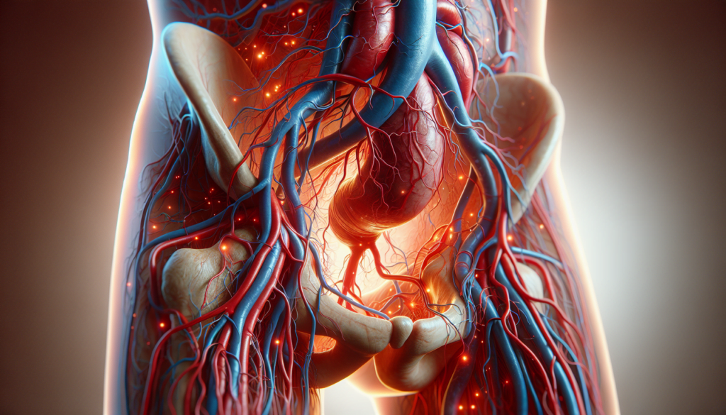
May-Thurner syndrome is a rare vascular condition that affects blood flow in the lower body. This disorder occurs when the left iliac vein is compressed by the right iliac artery, leading to an increased risk of deep vein thrombosis and other complications. Despite its potential severity, May-Thurner syndrome often goes undiagnosed, making awareness and understanding of this condition crucial for timely intervention and treatment.
This article delves into the key aspects of May-Thurner syndrome, providing valuable insights for patients and healthcare professionals alike. It explores the causes and risk factors associated with the condition, outlines common symptoms and potential complications, and discusses various treatment options available to manage the disorder effectively. By shedding light on this often-overlooked vascular issue, we aim to empower readers with the knowledge to recognize and address May-Thurner syndrome promptly.
Causes and Risk Factors


Anatomical Factors
May-Thurner syndrome develops due to a malfunction in the circulatory system, specifically between the right iliac artery and the left iliac vein. The right iliac artery brings oxygenated blood to the lower extremities, while the left iliac vein returns that blood to the heart for re-oxygenation. In some individuals, the right iliac artery compresses the left iliac vein against the lumbar spine, interrupting blood flow and potentially leading to venous stasis and thrombus formation. This anatomical variant has been shown to be present in over 20% of the population; however, not all individuals with this compression develop symptoms.
Certain anatomical factors such as the position of the right iliac artery and the width of the left iliac vein are thought to make the left iliac vein more susceptible to compression in patients with May-Thurner syndrome. Additionally, scoliosis can exacerbate the compression by altering the alignment of the spine and the overlying vessels.
Genetic Predisposition
While May-Thurner syndrome itself is not considered a hereditary condition, some studies suggest that there may be a genetic predisposition to developing the anatomical variant that leads to iliac vein compression. However, more research is needed to fully understand the potential role of genetics in the development of this condition.
Patients with May-Thurner syndrome often have a history of other venous disorders or a family history of blood clots, suggesting that there may be an underlying genetic component that increases the risk of developing venous issues. Certain inherited thrombophilic disorders, such as Factor V Leiden mutation and prothrombin gene mutation, can also increase the likelihood of developing deep vein thrombosis in patients with May-Thurner syndrome.
RELATED: Understanding Meningioma: Symptoms, Causes, and Treatments
Lifestyle and Environmental Factors
In addition to anatomical and genetic factors, certain lifestyle and environmental factors can increase the risk of developing symptoms related to May-Thurner syndrome. Prolonged periods of sitting or standing, such as during long flights or work shifts, can lead to venous stasis and exacerbate the effects of iliac vein compression. Dehydration can also contribute to the development of blood clots by increasing blood viscosity and reducing blood flow.
Women are more commonly affected by May-Thurner syndrome, particularly those who have given birth multiple times or those with a history of blood clots. Pregnancy itself can be a risk factor, as the growing uterus can exert pressure on the pelvic veins, further compromising blood flow in the presence of iliac vein compression. The use of oral contraceptives, which can increase the risk of blood clots, may also contribute to the development of symptoms in women with May-Thurner syndrome.
Symptoms and Complications
Early Warning Signs
May-Thurner syndrome often remains asymptomatic in its early stages, making it challenging to detect. However, some individuals may experience subtle signs and symptoms that can serve as early warning indicators of the condition. These initial symptoms may include a feeling of heaviness or discomfort in the left leg, particularly after prolonged periods of standing or sitting. Patients may also notice a slight swelling in the left leg, which can be more pronounced towards the end of the day. In some cases, individuals with May-Thurner syndrome may develop visible varicose veins on the left leg, a sign that blood flow is being compromised.
Advanced Symptoms
As May-Thurner syndrome progresses and the compression of the left iliac vein becomes more severe, patients may experience a range of advanced symptoms. One of the most common signs is significant swelling in the left leg, known as edema. This swelling can cause the leg to feel tight, heavy, and uncomfortable, making it difficult to move or perform daily activities. Patients may also experience pain or a throbbing sensation in the left leg, particularly when standing or walking for extended periods. In some cases, the skin on the affected leg may become discolored, taking on a reddish or purplish hue due to poor circulation. Advanced May-Thurner syndrome can also lead to the development of venous stasis dermatitis, a condition characterized by skin changes such as itching, dryness, and the formation of open sores or ulcers.
RELATED: Everything You Should Know About Mastoiditis: Symptoms and Solutions
Potential Complications
If left untreated, May-Thurner syndrome can give rise to serious complications that can have a significant impact on a patient’s health and quality of life. One of the most concerning complications is the development of deep vein thrombosis (DVT), a condition in which blood clots form within the deep veins of the leg. DVT can cause severe pain, swelling, and discoloration of the affected limb, and if the clot breaks free and travels to the lungs, it can result in a potentially life-threatening condition known as pulmonary embolism (PE). Patients with May-Thurner syndrome are at a higher risk of developing post-thrombotic syndrome (PTS), a chronic condition that occurs as a result of damage to the veins following a DVT. PTS can cause persistent pain, swelling, and skin changes in the affected leg, leading to long-term disability and reduced quality of life. In severe cases, May-Thurner syndrome can also contribute to the development of chronic venous insufficiency (CVI), a condition in which the veins struggle to efficiently return blood to the heart, leading to a range of symptoms and complications.
Treatment Options
Conservative Management
Conservative measures such as compression stockings and systemic anticoagulation are appropriate initial treatments for early signs of May-Thurner syndrome. These are aimed at preventing or decreasing the symptoms of post-thrombotic syndrome; however, conservative therapy alone is not adequate for long-term management due to its inability to address the underlying cause of venous compression. Patients with mild symptoms may benefit from a trial of conservative management, including modest weight gain for thin patients, elastic compression stockings for patients with pelvic or flank pain, and angiotensin inhibitors for patients with orthostatic proteinuria.
Minimally Invasive Procedures
The current standard of care for treatment of acute iliofemoral deep vein thrombosis in May-Thurner syndrome is urgent catheter-directed thrombolysis with mechanical thrombectomy, venoplasty, and common iliac venous stenting. This approach offers a minimally invasive option to actively treat both the mechanical compression with stent placement and the thrombus burden with chemical dissolution. Many authors advocate emergently placing a retrievable inferior vena cava filter via transjugular approach prior to endovascular thrombolysis in order to prevent embolization of the significant clot burden from the common iliac vein. Following thrombolysis, venoplasty with stent placement is the mainstay of endovascular treatment, typically performed with a large self-expanding nitinol or stainless-steel stent across the stenosis and into the inferior vena cava. Several studies have demonstrated long-term primary and secondary patency rates as high as 78% and 95%, respectively, at 2-year follow-up evaluation.
RELATED: Maladaptive Daydreaming: A Detailed Look at Symptoms and Solutions
Surgical Interventions
Several surgical decompression techniques have been described for May-Thurner syndrome, including saphenous vein bypass, placement of the right common iliac artery into a peritoneal sling, reimplantation of the left common iliac vein onto the vena cava, venotomy with excision of intraluminal adhesions, and vein patch venoplasty. Surgical treatment has an overall success rate, defined as patency of the left common iliac vein or venous bypass, of 40–88%. However, operative decompression is typically reserved as second-line treatment in cases where endovascular methods fail.
Conclusion
May-Thurner syndrome is a complex vascular condition that has a significant impact on blood flow in the lower body. This article has shed light on the causes, symptoms, and potential complications of this often-overlooked disorder. By exploring various treatment options, from conservative management to surgical interventions, we’ve aimed to provide a comprehensive overview to help patients and healthcare professionals better understand and address this condition.
Early detection and proper management are crucial to prevent the development of severe complications associated with May-Thurner syndrome. As medical knowledge and technology continue to advance, new and improved treatment methods are likely to emerge, offering hope for those affected by this condition. It’s essential for individuals experiencing symptoms to seek medical attention promptly and for healthcare providers to consider May-Thurner syndrome in their differential diagnosis, especially in cases of unexplained left leg swelling or recurrent deep vein thrombosis.

