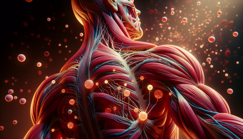
Myositis, a group of rare inflammatory muscle disorders, has a significant impact on the lives of those affected. This condition causes muscle inflammation and weakness, leading to various challenges in daily activities and overall quality of life. Understanding myositis is crucial for patients, caregivers, and healthcare professionals alike, as early diagnosis and proper management can make a big difference in outcomes.
This article aims to provide essential information about myositis, covering its causes, symptoms, and treatment options. It will explore the different types of myositis, discuss recent advances in diagnosis, and look into evolving treatment strategies. By shedding light on this complex condition, we hope to raise awareness and empower those dealing with myositis to make informed decisions about their health and care.
Myositis: An Overview
Myositis refers to a group of rare inflammatory muscle disorders characterized by muscle inflammation and weakness. The four main subtypes of idiopathic inflammatory myopathies (IIMs) are dermatomyositis (DM), polymyositis (PM), necrotizing myopathy (NM), and inclusion body myositis (IBM). The presence of autoantibodies and inflammatory infiltrates in the muscles suggests an autoimmune etiology, although the specific target autoantigens remain unidentified.
Definition and Classification
The classification of IIMs is based on clinical and histopathological features. The most commonly used criteria for PM and DM are the Peter/Bohan criteria, which include symmetric proximal muscle weakness, elevated serum muscle enzymes, myopathic changes on electromyography (EMG), characteristic muscle biopsy abnormalities, and the typical rash of DM.
RELATED: What You Need to Know About Premenstrual Dysphoric Disorder (PMDD)
Epidemiology of Myositis
The estimated prevalence of PM and DM ranges from 5 to 22 per 100,000 persons, with an incidence of approximately 1.2 to 19 per million persons at risk per year. DM has a bimodal age distribution, with peaks at 5-15 years and 45-60 years, while PM rarely occurs in children and has a mean age of onset between 50 and 60 years. Women are affected two to three times more often than men, and in the United States, the Black race to White race incidence ratio is 3 to 4:1.
Pathophysiology
The proposed pathogenetic mechanisms of myositis include:
- Direct effects of inflammatory cell infiltrates (CD4+ and CD8+ T cells, B cells, macrophages, and dendritic cells)
- Indirect effects of cytokines (interleukins, tumor necrosis factors, and interferons)
- Involvement of microvasculature (increased thickening of endothelial cells and expression of adhesion molecules)
- Humoral mechanisms (presence of autoantibodies in serum and complement and immunoglobulin in muscle biopsy specimens)
The histopathologic features of PM and DM include mononuclear inflammatory cell infiltrates, muscle fiber degeneration and regeneration, and perifascicular atrophy (a hallmark of DM). Immunopathology reveals differences between PM and DM, suggesting distinct pathogenic mechanisms, with T-cell-mediated muscle damage in PM and a role for microvessels in DM.
Clinical Presentation of Myositis
The clinical presentation of myositis can vary depending on the specific subtype and the individual patient. However, there are common muscular and extramuscular manifestations that characterize this group of disorders.
Muscular Symptoms
Muscle weakness is the hallmark feature of myositis. It typically affects the proximal muscles, those closest to the trunk, such as the shoulders, hips, and thighs. This weakness can make everyday tasks like climbing stairs, brushing hair, and getting in and out of cars challenging. The onset of weakness is usually gradual, occurring over weeks to months.
In addition to weakness, patients may experience muscle pain (myalgia) and tenderness. The affected muscles may also appear swollen due to inflammation. Patients often report a general feeling of being unwell, fatigue, weight loss, and night sweats.
RELATED: Understanding Ovarian Cysts: Symptoms, Causes, and Treatments
Extramuscular Manifestations
Dermatomyositis, a specific subtype of myositis, presents with distinctive skin manifestations in addition to muscle weakness. These include a reddish-purple rash (heliotrope rash) on the upper eyelids, face, neck, and backs of the hands, as well as swelling and discoloration around the eyes.
Myositis can also affect other organ systems. Some patients may experience difficulty swallowing (dysphagia) due to weakness of the esophageal muscles. Lung involvement, known as interstitial lung disease, can cause shortness of breath and a chronic cough. In rare cases, the heart muscle may be affected, leading to cardiac complications.
Disease Progression
The progression of myositis varies among individuals. Some may experience a single episode with complete recovery, while others have a more chronic, relapsing course. Inclusion body myositis tends to progress slowly over years, leading to severe muscle wasting and disability.
Prompt diagnosis and treatment are crucial for improving outcomes. If left untreated, myositis can lead to significant disability, with some patients becoming wheelchair-dependent. Complications such as aspiration pneumonia due to dysphagia and injuries from falls can have serious consequences.
Regular monitoring for extramuscular manifestations, particularly lung and heart involvement, is essential. With appropriate treatment and management, many patients with myositis can achieve remission and maintain a good quality of life.
Recent advances in myositis diagnosis have led to improved understanding and identification of the disease. Biomarkers and autoantibodies, imaging techniques, and histopathological findings all play crucial roles in diagnosing myositis accurately.
Myositis-specific autoantibodies (MSAs) and myositis-associated autoantibodies (MAAs) are important biomarkers that aid in diagnosis and classification of myositis subtypes. MSAs are highly specific for myositis, while MAAs are associated with overlap syndromes. Interferon signature genes and Janus kinase inhibition also show promise as diagnostic and therapeutic markers.
Imaging modalities such as MRI, ultrasound, and PET/CT have advanced myositis diagnosis. MRI can detect muscle inflammation, guide biopsy site selection, and monitor disease activity. Ultrasound is useful for detecting abnormal muscles and assessing treatment response. PET/CT identifies active inflammation and screens for associated malignancies.
Muscle biopsy remains the gold standard for myositis diagnosis. Histopathological findings like mononuclear cell infiltrates, muscle fiber necrosis, perifascicular atrophy, and rimmed vacuoles help differentiate myositis subtypes. Immunohistochemical analysis further enhances diagnostic accuracy.
Combining clinical presentation, autoantibody testing, imaging findings, and histopathology allows for a comprehensive diagnostic approach to myositis. These advances have significantly improved our ability to diagnose and manage this complex group of disorders.
Evolving Treatment Strategies
Traditional therapies for myositis include glucocorticoids and immunosuppressants. While effective in many patients, some have refractory disease or experience adverse effects. This has led to the exploration of novel treatment strategies.
RELATED: How to Recognize and Treat Obsessive Compulsive Disorder
Personalized medicine approaches aim to tailor treatment based on individual patient characteristics. Subgrouping patients by clinical phenotype, autoantibody status, and molecular signatures may help predict response to specific therapies.
Combining pharmacologic treatment with exercise has also shown benefits in muscle strength and quality of life. Future directions include further investigation of targeted biologics, optimizing combination regimens, and validating biomarkers to guide personalized treatment decisions in myositis.
Conclusion
Myositis remains a challenging condition, but recent advances in diagnosis and treatment offer hope for those affected. The combination of clinical assessment, biomarker testing, and advanced imaging techniques has improved our ability to identify and classify different types of myositis. This progress has a significant impact on patient care, allowing for more targeted and effective treatment strategies.
Looking ahead, the focus is on developing personalized approaches to manage myositis. Ongoing research into biological agents and the potential of combining pharmacological treatments with exercise therapy shows promise. As our understanding of myositis grows, so does the potential for better outcomes and an improved quality of life for patients living with this complex group of disorders.

