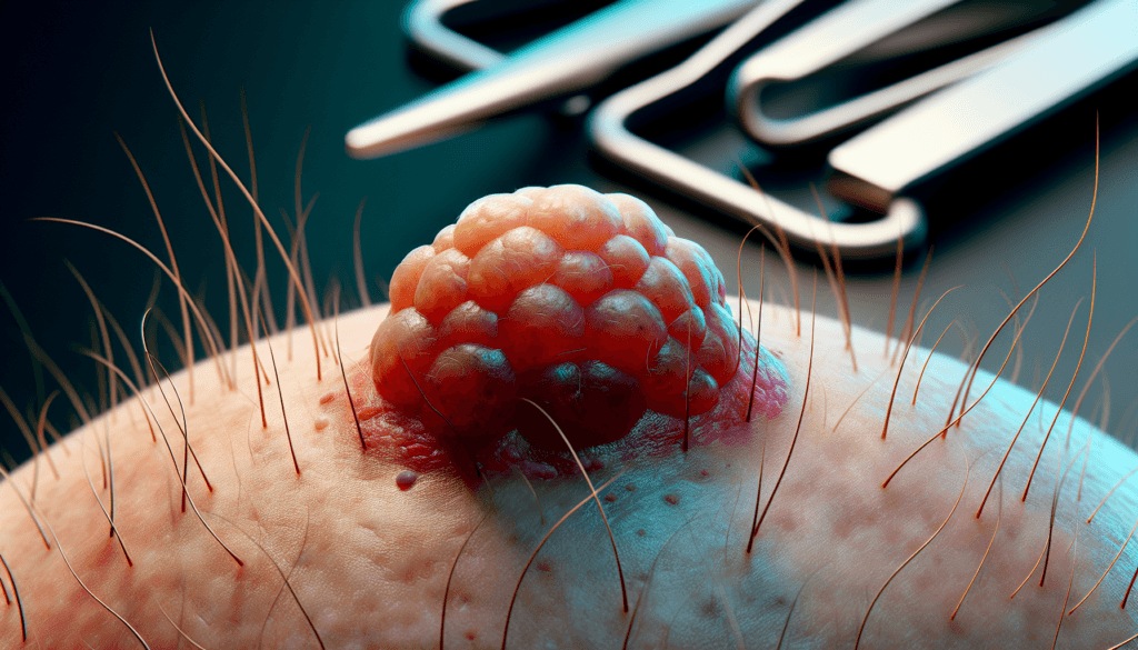
Pilar cysts, also known as trichilemmal cysts, are common benign growths that often appear on the scalp. These small, round bumps beneath the skin can cause concern for many individuals due to their visible nature and potential for discomfort. While typically harmless, pilar cysts can have an impact on a person’s appearance and self-confidence, leading many to seek information about their causes and treatment options.
Understanding pilar cysts is crucial for those affected by this condition. This article aims to provide a comprehensive overview of pilar cysts, including their symptoms, underlying causes, and available treatment methods. By exploring these aspects, readers will gain valuable insights to help them make informed decisions about managing their health and appearance. Whether dealing with a single cyst or multiple growths, this information will serve as a helpful guide to navigate the complexities of pilar cysts.
What is a Pilar Cyst?
Definition and Characteristics
Pilar cysts, also known as trichilemmal cysts, are noncancerous growths that arise from hair follicles. They are round, smooth, flesh-colored lumps that develop beneath the skin’s surface. The cysts are filled with keratin, a protein that makes up hair, skin, and nail cells. Pilar cysts tend to grow slowly over time, sometimes reaching a considerable size.
Common Locations
Pilar cysts most commonly occur on the scalp, where hair follicle density is highest. However, they can also appear on other parts of the body, such as the face, neck, arms, and legs. In some cases, multiple pilar cysts may develop simultaneously, especially on the scalp.
RELATED: How to Treat Meralgia Paresthetica: Expert Tips and Advice
Difference from Other Cyst Types
Pilar cysts are often confused with sebaceous cysts, but there are distinct differences between the two. While pilar cysts originate from hair follicles, sebaceous cysts develop from sebaceous glands, which secrete an oily substance called sebum. Additionally, the contents of a pilar cyst primarily consist of keratin, whereas sebaceous cysts contain sebum. Pilar cysts are typically firmer and more mobile than sebaceous cysts. It is important to note that pilar cysts are generally benign and do not pose a significant health risk. However, in rare cases (approximately 3%), pilar cysts can transform into proliferating trichilemmal tumors, which are characterized by rapid growth and potential ulceration. These tumors may require more extensive treatment, such as surgical excision, to prevent further complications.
Symptoms and Identification
Physical Appearance
Pilar cysts typically appear as smooth, round, flesh-colored lumps beneath the skin’s surface. They are usually firm to the touch and can be easily moved under the skin. The size of pilar cysts can vary, ranging from small, pea-sized bumps to larger growths that may reach several centimeters in diameter. In some cases, multiple pilar cysts may develop in close proximity to one another, especially on the scalp.
Associated Discomfort
In most cases, pilar cysts are painless and do not cause significant discomfort. However, if a cyst becomes inflamed or infected, it may become tender, red, and swollen. This can occur if the cyst ruptures, either spontaneously or due to trauma, allowing bacteria to enter the affected area. Additionally, larger pilar cysts may cause discomfort if they press on underlying structures, such as nerves or blood vessels.
When to Seek Medical Attention
While pilar cysts are generally benign, it is important to seek medical attention if any of the following symptoms occur:
- Rapid growth or changes in the appearance of the cyst
- Pain, redness, or swelling around the cyst
- Drainage of pus or other fluids from the cyst
- Ulceration or bleeding from the cyst
- Recurrence of previously removed cysts
These symptoms may indicate an infection, inflammation, or, in rare cases, malignant transformation. A healthcare provider can properly evaluate the cyst and recommend appropriate treatment options. Additionally, individuals who are bothered by the appearance of pilar cysts or experience discomfort due to their presence may wish to consult with a dermatologist to discuss removal options for cosmetic purposes.
Causes and Risk Factors
Keratin Buildup
Pilar cysts develop when keratin, a protein that makes up hair, skin, and nail cells, accumulates in the lining of hair follicles. This buildup occurs gradually over time, causing the formation of smooth, round lumps beneath the skin’s surface. As the keratin continues to accumulate, the cyst may grow larger, sometimes reaching a considerable size.
Genetic Predisposition
Genetic factors play a significant role in the development of pilar cysts. In many cases, individuals with a family history of these cysts are more likely to develop them. The tendency to form pilar cysts can be inherited in an autosomal dominant pattern, meaning that if one parent carries the genetic trait, their children have a 50% chance of inheriting it and developing cysts.
RELATED: A Detailed Guide to Meibomian Gland Dysfunction and Its Treatment
Age and Gender Considerations
While pilar cysts can occur in individuals of any age, they are most commonly observed in middle-aged adults. Women are more frequently affected by these cysts than men, although the exact reason for this gender disparity remains unclear. Hormonal factors may contribute to the higher incidence of pilar cysts in women, but further research is needed to confirm this hypothesis.
It is important to note that while genetic predisposition and age are significant risk factors for developing pilar cysts, not everyone with a family history or in the middle-age range will necessarily develop these growths. Additionally, individuals without a known family history may still develop pilar cysts, suggesting that other factors, such as environmental influences or random genetic mutations, may also play a role in their formation.
Diagnosis and Treatment Options
Medical Examination Process
The diagnosis of pilar cysts typically involves a thorough physical examination by a healthcare provider. During the examination, the provider will assess the appearance, size, and location of the cyst. In some cases, imaging studies such as computed tomography (CT) scans or magnetic resonance imaging (MRI) may be ordered to evaluate the extent of the cyst’s growth and to determine if it has invaded the underlying bone or soft tissue.
Surgical Removal Techniques
The primary treatment for pilar cysts is surgical excision. The procedure involves numbing the affected area with local anesthesia and making a small incision to remove the entire cyst, including its wall or sac. Complete removal of the cyst wall is crucial to prevent recurrence. After the cyst is removed, the incision is closed with sutures, and the tissue may be sent for pathological examination to confirm the diagnosis and rule out any malignant transformation.
In rare cases of proliferating trichilemmal cysts, which have a higher risk of malignancy, more extensive surgical interventions may be necessary. These cases might require multiple surgical sessions, radiation therapy, or chemotherapy to manage the aggressive growth and potential spread of the tumor.
RELATED: Navigating Myopathy: From Diagnosis to Treatment Solutions
Non-Surgical Management
While surgical excision is the most effective treatment for pilar cysts, there are some non-surgical management options that may be considered in certain situations. If a cyst becomes inflamed or infected, it is generally not recommended to remove it surgically until the inflammation subsides. In such cases, wound swab and culture sensitivity tests can be performed to identify any infection and guide appropriate treatment with antibiotics.
Conservative management of pilar cysts may include warm compresses to help reduce inflammation and encourage drainage. Over-the-counter pain medications can be used to alleviate discomfort associated with inflamed cysts. However, it is important to note that non-surgical approaches do not eliminate the cyst and may only provide temporary relief of symptoms.
Conclusion
Pilar cysts, while generally harmless, can have an impact on a person’s appearance and comfort. This article has provided a comprehensive look at these common growths, covering their characteristics, causes, and treatment options. Understanding the nature of pilar cysts and their potential complications is crucial to make informed decisions about managing them.
For those dealing with pilar cysts, various approaches are available to address them. While surgical removal remains the most effective method to eliminate these growths, non-surgical options can help manage symptoms in certain cases. Ultimately, consulting with a healthcare provider is essential to determine the best course of action based on individual circumstances and preferences.

