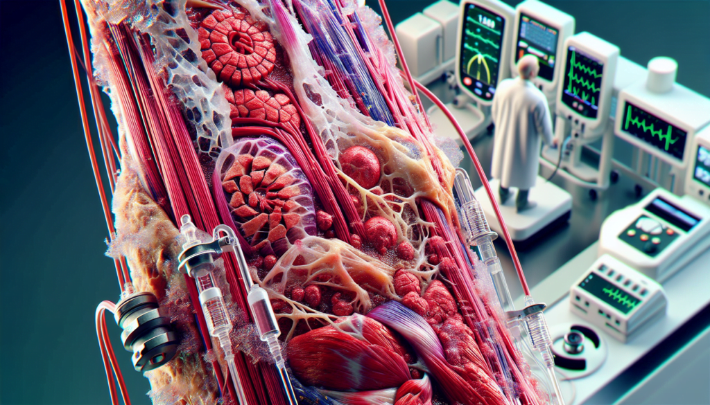
Rhabdomyolysis is a serious medical condition that can have severe consequences if left untreated. This muscle breakdown disorder occurs when damaged skeletal muscle tissue releases its contents into the bloodstream, potentially leading to kidney damage and other complications. Understanding rhabdomyolysis is crucial for early detection and prompt treatment, as it can affect anyone from athletes to individuals with certain medical conditions.
This article delves into the causes, symptoms, and treatment options for rhabdomyolysis. It explores common triggers, such as intense physical exertion and certain medications, and highlights key warning signs to watch out for. Additionally, it discusses diagnostic procedures and treatment approaches, providing valuable information to help readers recognize and respond to this potentially life-threatening condition.
What is Rhabdomyolysis?


Rhabdomyolysis is a complex medical condition involving the rapid dissolution of damaged or injured skeletal muscle. This disruption of skeletal muscle integrity leads to the direct release of intracellular muscle components, including myoglobin, creatine kinase (CK), aldolase, and lactate dehydrogenase, as well as electrolytes, into the bloodstream and extracellular space.
Definition
The word rhabdomyolysis is derived from the Greek words rhabdos (rod-like/striated), mus (muscle), and Lucis (breakdown). It ranges from an asymptomatic illness with elevation in the CK level to a life-threatening condition associated with extreme elevations in CK, electrolyte imbalances, acute renal failure (ARF), and disseminated intravascular coagulation.
Pathophysiology
The final common pathway leading to rhabdomyolysis involves either direct myocyte injury or a failure of the energy supply within the muscle cells. This disruption causes an excessive intracellular influx of Na+ and Ca2+, leading to a sustained myofibrillar contraction that further depletes ATP. The elevation in Ca2+ activates Ca2+-dependent proteases and phospholipases, promoting lysis of the cellular membrane and further damage to the ion channels. The end result is an inflammatory, self-sustaining myolytic cascade that causes necrosis of the muscle fibers and releases the muscle contents into the extracellular space and the bloodstream.
Prevalence
Approximately 25,000 cases of rhabdomyolysis are reported each year in the USA. The prevalence of acute kidney injury in rhabdomyolysis is about 5 to 30%. Males, African-American race, obesity, age more than 60 are factors that demonstrate a higher incidence of rhabdomyolysis. Even without acute kidney injury, the mortality rate is about 20%, and with kidney injury, mortality is about 50%.
Common Causes of Rhabdomyolysis
Rhabdomyolysis has a wide range of causes, from physical trauma to medical conditions. Some of the most common causes include:
Trauma and Crush Injuries
Blunt injuries and crush injuries are frequent causes of trauma-induced rhabdomyolysis. Interestingly, in crush injuries associated with severe natural disasters or man-made traumatic events such as bombings, earthquakes, or building collapses, the onset of rhabdomyolysis is noted to occur only once the acute compression of muscle is relieved, thereby allowing the products of muscle breakdown to enter the circulatory system. High-voltage electrical injuries (i.e., electrocution or lightning strikes) are other causes of trauma-induced rhabdomyolysis. Up to 10% of patients who survive the initial electrical accident are estimated to develop rhabdomyolysis.
RELATED: Understanding Multiple Sclerosis: Symptoms, Causes, and Treatments
Extreme Physical Exertion
Exertional or exercise-induced rhabdomyolysis is a condition caused by unaccustomed physical exercise and characterized by a breakdown of skeletal muscles that leads to the release of its intracellular components, such as myoglobin and creatine kinase (CK), into the circulatory system. It can cause severe complications, including acute kidney injury (AKI), disseminated intravascular coagulation (DIC), compartment syndrome, cardiac arrhythmia, liver dysfunction, and various electrolyte derangements such as hypocalcemia, hypercalcemia, hyperkalemia, hyperphosphatemia, hypomagnesemia, and hyperuricemia, along with mortality risk.
One of the major challenges in diagnosing exertional rhabdomyolysis is the fact that serum CK levels will naturally rise after strenuous exercise in almost all normal humans, potentially up to 10 times the higher limit of normal. The increase in CK levels also varies widely among patients, and it is possible for one individual to develop exertional rhabdomyolysis while exerting the same energy under the same conditions as another individual who does not develop exertional rhabdomyolysis. Increased temperature and humidity during exercise/exertion may also play a role in higher rates of rhabdomyolysis.
Medications and Toxins
Drug toxicity involves organs like the kidneys, liver, gastrointestinal tract, and central nervous system (CNS), as these are the ones that are most impacted by both medications and drugs of abuse. Drug-induced rhabdomyolysis can be the result of direct injury to the muscle caused by myotoxic drugs or intravenous drug use, as well as secondary muscle injuries such as inadequate blood supply or injury caused by prolonged seizures. Rhabdo may also occur in cases where the person lost consciousness or overdosed and fell and injured or crushed a muscle.
Drugs that cause rhabdomyolysis include:
- Antipsychotics and antidepressants: Amoxapine, Lithium, Protriptyline, Phenelzine, Chlorpromazine, Loxapine, Promazine, Trifluoperazine
- Sedative hypnotics: Benzodiazepines, Nitrazepam, Triazolam, Barbiturates.
- Antilipemic agents: Lovastatin, Pravastatin, Simvastatin, Clozafibrate, Ciprofibrate, Clofibrate
- Drugs of Abuse: Heroin, Cocaine, Methadone, D-lysergic acid diethylamide (LSD)
- Antihistamines: Diphenhydramine, Doxylamine
Medical Conditions
Various medical conditions can also lead to rhabdomyolysis, such as:
- Infections: Rhabdomyolysis has been described in all types of infections, ranging from localized muscle infections with erythema (bacterial pyomyositis) to patients with sepsis and no direct muscle infection. Proposed mechanisms for the development of rhabdomyolysis include tissue hypoxia secondary to sepsis or dehydration, toxin release, associated fever, direct bacterial invasion of muscle, or rigors/tremors. Classically, Legionella bacteria have been associated with bacterial rhabdomyolysis. Viral infections have also been implicated in rhabdomyolysis development, most commonly influenza A and B viruses. Rhabdomyolysis because of other viruses such as HIV, coxsackie virus, Epstein-Barr virus, Cytomegalovirus, herpes simplex virus, varicella zoster virus, and West Nile virus has also been described.
- Muscle Ischemia: Muscle cell necrosis can result from prolonged periods of oxygen deprivation to muscle, ultimately precipitating rhabdomyolysis and ARF. Causes of localized muscle ischemia include compression of blood vessels during surgery or otherwise, thromboses, emboli, compartment syndrome, carboxyhemoglobinemia, or sickle cell disease. Although rare, hypothermia can lead to the development of rhabdomyolysis by reducing muscle perfusion.
- Genetic Disorders: Genetic polymorphisms and defects accounting for skeletal muscle diseases potentiate the risk for episodes of rhabdomyolysis. These defects include enzymes from the glycolysis and glycogenolysis pathway and pentose phosphate pathway. Impaired mitochondrial pathways involve fatty acid oxidation, the citric acid cycle and the mitochondrial respiratory chain. And finally, defects in the Ca2+ homeostasis are seen in patients with mutations in proteins involved in excitation-contraction coupling, myotonias and skeletal muscle dystrophies.
Recognizing Symptoms of Rhabdomyolysis
The classic triad of symptoms associated with rhabdomyolysis includes muscle pain, weakness, and dark-colored urine. However, this triad is observed in less than 10% of patients. More than 50% of patients do not complain of muscle pain or weakness, with the initial presenting symptom being discolored urine.
Muscle pain, when present, is typically most prominent in proximal muscle groups, such as the thighs, shoulders, lower back, and calves. The pain may be accompanied by stiffness and cramping. Muscle weakness usually affects the same muscle groups as the pain, with the proximal legs being most frequently involved.
Muscle swelling affects 8 to 52% of patients with rhabdomyolysis. When it occurs, detectable swelling in the extremities generally develops with fluid repletion. Swelling may be due to either myoedema, which is nonpitting and apparent at presentation or develops after rehydration, or peripheral edema, which is pitting and occurs with rehydration, particularly in patients with acute kidney injury.
Dark-colored urine, ranging from red to brown or “tea-colored” to “cola-colored,” is one of the classic signs of rhabdomyolysis but occurs in 10% or fewer cases. Myoglobin, a heme-containing respiratory protein released from damaged muscle, is rapidly excreted in the urine, often resulting in red to brown urine. Visible changes in urine color occur once urine myoglobin levels exceed approximately 100 to 300 mg/dL.
Other Common Symptoms
In addition to the classic triad, patients with rhabdomyolysis may experience:
- Malaise
- Fever
- Tachycardia
- Nausea and vomiting
- Abdominal pain
These symptoms are more common in severely affected patients.
RELATED: All About Moles: From Causes to Treatment Options
When to Seek Medical Attention
Rhabdomyolysis should be suspected in patients presenting with any of the following:
- The classic triad of muscle pain, weakness, and dark-colored urine
- A potential cause, triggering event, or increased risk of rhabdomyolysis, with or without myalgias or pigmenturia, as symptoms may be vague or absent in up to 50% of patients
- Acute muscle weakness and marked elevation of creatine kinase (CK)
Prompt recognition and treatment are crucial to prevent severe complications such as acute kidney injury, compartment syndrome, and disseminated intravascular coagulation. If rhabdomyolysis is suspected, immediate medical attention should be sought for proper diagnosis and management.
Diagnosis and Treatment
Rhabdomyolysis should be suspected in patients presenting with any of the following:
- The classic triad of muscle pain, weakness, and dark-colored urine
- A potential cause, triggering event, or increased risk of rhabdomyolysis, with or without myalgias or pigmenturia, as symptoms may be vague or absent in up to 50% of patients
- Acute muscle weakness and marked elevation of creatine kinase (CK)
Prompt recognition and treatment are crucial to prevent severe complications such as acute kidney injury, compartment syndrome, and disseminated intravascular coagulation. If rhabdomyolysis is suspected, immediate medical attention should be sought for proper diagnosis and management.
Diagnostic Tests
Useful laboratory tests that should be ordered include:
- Complete blood count (CBC), including hemoglobin, hematocrit, and platelets
- Serum chemistries, including blood urea nitrogen (BUN), creatinine, glucose, calcium, potassium, phosphate, uric acid, and liver function tests (LFTs)
- Prothrombin time (PT) and activated partial thromboplastin time (aPTT)
- Serum aldolase and lactate dehydrogenase (LDH)
The most reliable and sensitive indicator of muscle injury is creatine kinase (CK). CK levels rise within 12 hours of muscle injury, peak in 24-36 hours, and decrease at a rate of 30-40% per day. CK levels 5 times the reference range suggest rhabdomyolysis, though levels are frequently as high as 100 times the reference range or even higher.
Urine myoglobin measurements are preferable to plasma myoglobin due to the short half-life of myoglobin. A urine dipstick test for blood that has positive findings in the absence of red blood cells (RBCs) suggests myoglobinuria. However, a normal test result does not rule out rhabdomyolysis.
Treatment Approaches
The goal of rhabdomyolysis management is to maintain adequate fluid resuscitation and prevent acute kidney injury. Identifying the underlying cause and its removal is the first step in managing patients. Management should include:
- Continuous assessment of airway, breathing, and circulation
- Frequent examinations
- Appropriate hydration to improve end-organ perfusion
- Close monitoring of urine output
- Correction of electrolyte abnormalities
- Identification of complications like compartment syndrome and disseminated intravascular coagulation
In patients with crush injury, the initiation of IV hydration/fluid resuscitation should begin as early as possible at the site of injury, even before relieving injury if possible. A liberal amount of fluids should be given up to 10 to 20 L to maintain adequate intravascular volume and diuresis.
Alkalization of urine is believed to be helpful and is based on the observation that acidic urine is necessary to cause acute tubular necrosis. A suggested regimen for adult patients is isotonic sodium chloride solution (0.9% NaCl) with 1 ampule of sodium bicarbonate administered at 100 mL/h. The goal of alkaline fluid infusion is to maintain a serum pH not to exceed 7.5 and a urine pH just above 6.5.
RELATED: Myalgic Encephalomyelitis: Symptoms, Causes, and Treatments
Complications Management
Frequently monitor serum electrolyte levels, urine pH levels, and acid-base status. Treatment of hyperkalemia consists of IV sodium bicarbonate, glucose, and insulin; oral or rectal sodium polystyrene sulfonate; and hemodialysis. Administer IV calcium chloride for patients who are hemodynamically compromised and hyperkalemic.
Hypocalcemia is noted early in the course of rhabdomyolysis and generally is not of clinical significance. Correct hypocalcemia only if the patient has cardiac dysrhythmias or seizures. Calcium supplementation is not recommended, as hypercalcemia may be seen in the recovery phase.
Dialysis may be required in patients with oliguric renal failure, persistent hyperkalemia, other electrolyte abnormalities, pulmonary edema, congestive heart failure, and persistent metabolic acidosis.
Surgical care may be necessary, depending on the cause of rhabdomyolysis. Compartment pressures should be measured when significant muscle injury has occurred. When the intracompartmental pressure exceeds 30 mm Hg, a fasciotomy is advocated. Limb fractures may require surgical and orthopedic treatment.
Conclusion
Rhabdomyolysis is a serious condition that can have a significant impact on a person’s health. Understanding its causes, symptoms, and treatment options is crucial to ensure prompt medical attention. This knowledge empowers individuals to recognize potential warning signs and seek help quickly, potentially preventing severe complications like kidney damage. The diverse range of triggers, from intense physical activity to certain medications, highlights the importance of staying aware and informed about this condition.
Proper diagnosis and treatment of rhabdomyolysis are key to managing its effects and promoting recovery. Healthcare professionals use a combination of clinical assessment and lab tests to identify rhabdomyolysis and start appropriate treatment. The focus on maintaining adequate hydration and addressing electrolyte imbalances underscores the complexity of managing this condition. By staying informed and acting quickly when symptoms arise, individuals can play a vital role in their own health and well-being when it comes to rhabdomyolysis.

