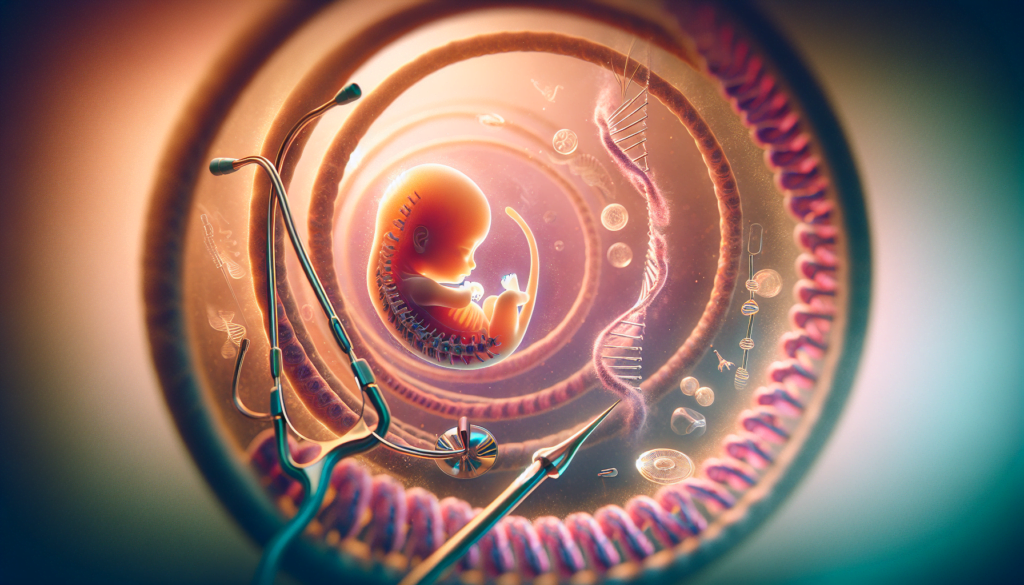
Spina bifida is a complex birth defect that affects the spinal cord and surrounding structures. This condition occurs when the neural tube, which eventually forms the brain and spinal cord, doesn’t close properly during early fetal development. The impact of spina bifida can range from mild to severe, depending on the location and extent of the spinal defect.
Living with spina bifida presents unique challenges at every stage of life. This article delves into the causes, symptoms, and various treatment options available for individuals with spina bifida. We’ll explore the diagnostic process, examine the spectrum of treatments, and discuss how spina bifida affects people from infancy through adulthood. By understanding this condition better, we aim to provide valuable insights for those affected by spina bifida and their caregivers.
Spina Bifida: A Multifaceted Condition


Spina bifida is a complex birth defect that occurs when the spine and spinal cord do not form properly during early fetal development. The condition can range from mild to severe, depending on the location and extent of the spinal defect. Spina bifida has an influence on various aspects of an individual’s life, including physical mobility, bladder and bowel function, and cognitive development.
Embryonic Development
During the first month of pregnancy, the neural tube, which eventually forms the brain and spinal cord, typically closes by the 28th day after conception. In babies with spina bifida, a portion of the neural tube fails to close completely, leading to the development of the condition. The incomplete closure of the neural tube can occur at different locations along the spine, resulting in varying degrees of severity.
Classification of Types
Spina bifida is classified into three main types:
- Spina bifida occulta: The mildest and most common form, often causing no symptoms and requiring no treatment. The skin covers the deformity of the spinal bone, and the spinal cord is not affected.
- Meningocele: A rare type where the meninges (membrane surrounding the spinal cord) protrude through an opening in the spine, forming a lump or sac on the back. Surgery can repair the defect with little or no nerve damage.
- Myelomeningocele: The most severe form, occurring when part of the spinal cord and meninges emerge through an opening in the spine. This type can cause significant nerve damage, leading to paralysis, bladder and bowel dysfunction, and cognitive issues.
RELATED: Smegma: Practical Steps for Cleaning and Prevention
Associated Conditions
Individuals with spina bifida, particularly those with myelomeningocele, may experience various associated conditions:
- Hydrocephalus: A buildup of excess fluid in the brain, often requiring surgical treatment with a shunt to drain the fluid and prevent brain damage.
- Chiari malformation (Type II): A condition where brain tissue extends into the spinal canal, potentially causing pressure on the spinal cord or brainstem.
- Tethered spinal cord: The spinal cord becomes stuck to the lining of the spinal canal, leading to reduced sensitivity, bladder problems, and orthopedic issues.
- Orthopedic conditions: Curved spine (scoliosis), clubfoot, hip dislocation, and muscle contractures.
- Latex allergy: Many individuals with spina bifida have a higher risk of an allergic reaction to natural rubber or latex products.
Incidence and Demographics
In the United States, spina bifida occurs in about 1 in every 2,875 births. The condition affects people of all racial and ethnic backgrounds, but there are some notable differences in prevalence:
| Racial/Ethnic Group | Prevalence per 10,000 Live Births |
|---|---|
| Hispanic | 3.80 |
| Non-Hispanic white | 3.09 |
| Non-Hispanic black | 2.73 |
Prevention efforts, such as ensuring adequate folic acid intake before and during early pregnancy, have been shown to reduce the risk of spina bifida. Since the introduction of folic acid fortification in grain products in the United States, there has been a 28% reduction in the prevalence of neural tube defects, including spina bifida.
Diagnostic Journey
The diagnostic journey for spina bifida begins before the baby is born. Prenatal screening tests can check for spina bifida and other congenital anomalies. These tests are not perfect, but they provide valuable information to help prepare for the baby’s arrival and any necessary treatments.
Maternal Serum Screening
One of the first steps in diagnosing spina bifida is the maternal serum alpha-fetoprotein (MSAFP) test. This blood test, typically performed between 16 and 18 weeks of pregnancy, measures the level of alpha-fetoprotein (AFP) in the mother’s blood. Abnormally high levels of AFP may indicate that the fetus has spina bifida or another neural tube defect. However, high AFP levels can also be caused by other factors, such as an incorrect estimate of the fetus’s age or the presence of multiple fetuses. If AFP levels are high, additional testing is necessary to confirm the diagnosis.
RELATED: Everything You Need to Know About Skin Tags
Detailed Ultrasound
An ultrasound exam is the most accurate way to diagnose spina bifida before delivery. During pregnancy, an ultrasound may be performed in the first trimester (11 to 14 weeks) or the second trimester (18 to 22 weeks). The second-trimester ultrasound is crucial for identifying and ruling out conditions that may be present at birth. An advanced ultrasound can detect symptoms of spina bifida, such as an open spine or features in the baby’s brain. The ultrasound can also help determine the severity of the condition.
Amniocentesis and Other Tests
If the prenatal ultrasound confirms the diagnosis of spina bifida, the healthcare provider may recommend amniocentesis. During this test, a small sample of amniotic fluid is removed from the sac surrounding the baby. The fluid is tested for high levels of AFP and other indicators of spina bifida. Amniocentesis carries a slight risk of pregnancy loss, so it is important to discuss the potential risks and benefits with a healthcare professional.
In some cases, additional tests may be performed to gather more information about the baby’s condition. These tests may include:
- Fetal MRI: This imaging test provides detailed images of the baby’s brain and spinal cord, helping to identify the type of spina bifida and any associated complications, such as hydrocephalus or Chiari malformation.
- Quadruple screen test: This blood test, performed between 15 and 22 weeks of pregnancy, measures levels of four substances produced during pregnancy. Elevated levels of these substances may indicate an increased risk of spina bifida or other neural tube defects.
Postnatal Confirmation
While spina bifida is often diagnosed before birth, some cases may not be detected until after the baby is born. In these instances, physical examination of the newborn can reveal signs of spina bifida, such as a hairy patch, dimple, dark spot, or swelling on the baby’s back at the site of the spinal malformation. Imaging tests, such as X-rays, MRI, or CT scans, can provide a clearer view of the baby’s spine and confirm the diagnosis.
Throughout the diagnostic journey, it is essential for parents to work closely with their healthcare team, including obstetricians, pediatric neurosurgeons, and other specialists, to ensure the best possible care for their baby. Early diagn
Treatment Spectrum
The treatment spectrum for spina bifida encompasses a range of interventions, from prenatal care to lifelong management. Early diagnosis and treatment are crucial to optimize outcomes and quality of life for individuals with spina bifida.
Prenatal Interventions: Prenatal surgery for myelomeningocele, the most severe form of spina bifida, has shown clear benefits compared to standard postnatal treatment. The Management of Myelomeningocele Study (MOMS) found that fetal surgery decreased the risk of death or need for a shunt to alleviate fluid build-up in the brain by 12 months of age and improved scores on developmental testing for both mental and motor function at 30 months of age. Fetal surgery involves covering the myelomeningocele with multiple layers of the fetus’s own tissue during pregnancy, offering a rare opportunity to improve outcomes for the developing baby.
Neonatal Care: Immediate care after birth is critical for babies with spina bifida. Surgery to close the defect is usually performed within the first 48 hours to preserve neural tissue and prevent infection. Careful handling of the baby is essential to protect the exposed spinal cord, which may include using a protective device and special positioning. Antibiotics are given to prevent infections, particularly in the meninges and urinary tract.
Surgical Management:
- Spinal cord surgery: For babies with myelomeningocele or meningocele, neurosurgery may be needed within the first few days after birth to place the protruding spinal cord in the spinal column, reconstruct tissues, and close the gap in the skin.
- Hydrocephalus treatment: About 80% of babies with spina bifida develop hydrocephalus after surgery. A second operation may be needed to place a ventricular peritoneal (VP) shunt, a small drainage tube that redirects extra fluid from the brain.
- Chiari malformation treatment: Some babies with spina bifida may have a Chiari malformation, where part of the brain pushes into the spinal canal. Neurosurgery to reduce pressure on the spinal cord or brainstem may be recommended.
- Tethered spinal cord treatment: If the spinal cord remains attached to tissue near the lower spine, causing symptoms such as pain or bladder problems, surgery to release the spinal cord may be necessary.
Multidisciplinary Approach: Ongoing care from a multidisciplinary team of specialists is essential for individuals with spina bifida. This team may include:
- Pediatric neurosurgery
- Orthopedic surgery
- Urology
- Physical medicine and rehabilitation
- Social work
- Physical/occupational therapy
The multidisciplinary approach ensures optimal and individualized care, addressing the complex needs of patients with spina bifida. Regular communication between clinicians and vigilance on the part of families are key to identifying and treating complications promptly. As children with spina bifida grow, seamless transition to multidisciplinary clinics ensures continued comprehensive care throughout their lives.
Life Stages with Spina Bifida
Living with spina bifida presents unique challenges at every stage of life. From infancy through adulthood, individuals with this condition and their families navigate a complex journey that requires ongoing care, support, and adaptability.
Infancy and Early Childhood
In the early years, care for infants with spina bifida focuses on treating problems that can worsen their condition and identifying changes in their health status. Parents and caregivers learn about the condition and how to meet their child’s special needs, such as performing catheterization and protecting the skin from sores and injuries.
Physical therapy plays a crucial role in helping babies with spina bifida develop strength and flexibility in their legs. Occupational therapy assists with basic dressing and grooming skills, while braces and other orthotic devices support proper leg development and mobility.
School-Age Challenges
As children with spina bifida enter school, they may face various challenges related to their condition. Some may experience learning difficulties, particularly those with hydrocephalus managed by shunts. Attention problems, slow work pace, and organizational issues are common concerns.
Individualized Education Plans (IEPs) and 504 Plans are essential tools for ensuring that children with spina bifida receive the accommodations and support they need to succeed academically. These plans may include provisions for assistive technology, wheelchair accessibility, and additional time for assignments.
Encouraging independence is a key focus during this stage. Children with spina bifida should be given opportunities to take responsibility for self-care tasks, make choices, and engage in physical activities adapted to their abilities.
Adolescence and Transition
The teenage years bring new challenges as adolescents with spina bifida transition from pediatric to adult health care systems. This period also marks a shift towards greater independence in managing their condition and making decisions about their future.
Teens with spina bifida may struggle with self-esteem, social relationships, and the emotional impact of their disability. Support from family, friends, and healthcare professionals is crucial during this time.
Transition planning should begin early to ensure a smooth shift to adult healthcare providers and to address issues such as education, employment, and independent living. Vocational rehabilitation services and independent living skills courses can help teens prepare for adulthood.
RELATED: Shin Splints: Essential Information on Symptoms and Treatment
Adulthood and Aging
As individuals with spina bifida reach adulthood, they may encounter new health challenges related to aging with their condition. These can include urinary tract infections, pressure sores, respiratory problems, and orthopedic issues such as scoliosis and joint deterioration.
Regular check-ups with a multidisciplinary healthcare team are essential for monitoring and managing these concerns. Adults with spina bifida should also maintain a healthy lifestyle, including a balanced diet, regular exercise, and stress management techniques.
In addition to health concerns, adults with spina bifida may face barriers in education, employment, relationships, and community participation. Advocacy efforts aim to promote accessibility, inclusion, and equal opportunities for individuals with disabilities.
Throughout all stages of life, ongoing research offers hope for improved treatments and outcomes for those living with spina bifida. By staying informed, connected to support networks, and proactive in their care, individuals with this condition can lead fulfilling lives and reach their full potential.
Conclusion
Spina bifida has a significant impact on individuals throughout their lives, from prenatal diagnosis to adulthood. The condition presents unique challenges at each stage, requiring ongoing medical care, support, and adaptability. Advances in prenatal surgery, multidisciplinary care approaches, and improved understanding of the condition have led to better outcomes and quality of life for those affected.
As research continues, there’s hope for new treatments and improved management strategies to address the complex needs of individuals with spina bifida. By fostering awareness, promoting inclusive environments, and providing comprehensive support, we can empower those living with spina bifida to reach their full potential. The journey with spina bifida is ongoing, but with proper care and support, individuals can lead fulfilling lives and overcome the challenges they face.

