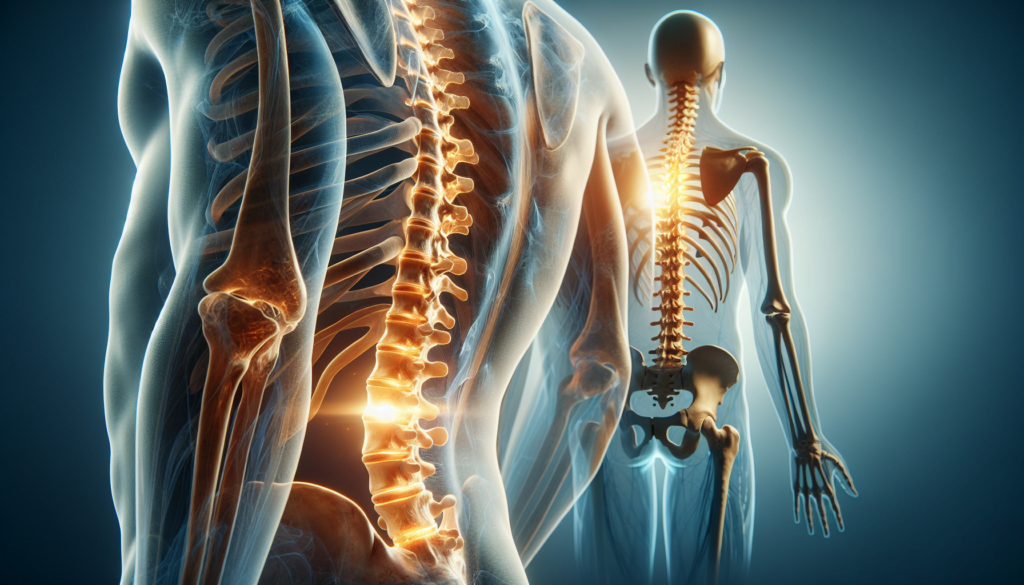
Spondylolisthesis is a spinal condition that can cause significant discomfort and impact daily life. This occurs when one vertebra slips forward over the one below it, leading to potential nerve compression and back pain. Understanding this condition is crucial for those affected, as it can have an influence on mobility and overall quality of life.
This article aims to provide a comprehensive overview of spondylolisthesis. It will explore the anatomy of the spine, delve into the causes and risk factors associated with the condition, and discuss its clinical presentation. Additionally, it will examine various treatment approaches available to manage spondylolisthesis, from conservative methods to surgical interventions. By the end, readers will have a clearer understanding of this spinal disorder and the options to address it.
Anatomy of the Spine
The human spine is a complex structure that provides support, flexibility, and protection to the spinal cord. It consists of 33 vertebrae stacked on top of each other, separated by intervertebral discs. The spine has an S-shaped curve when viewed from the side, which helps to distribute weight evenly and absorb shock.
Vertebral Structure
Each vertebra has a cylindrical body in the front and a vertebral arch in the back. The arch is made up of two pedicles and two laminae, which form the spinal canal that houses the spinal cord. Between each vertebra, there are small joints called facet joints that allow for movement and stability.
The intervertebral discs sit between the vertebral bodies and act as shock absorbers. They have a tough outer layer called the annulus fibrosus and a soft, gel-like center called the nucleus pulposus. These discs help to maintain the spine’s flexibility and prevent the vertebrae from rubbing against each other.
RELATED: Astrovirus Explained: From Symptoms to Treatment
Spinal Stability
Spinal stability is maintained by a combination of bony structures, ligaments, and muscles. The facet joints and intervertebral discs play a crucial role in keeping the vertebrae aligned and preventing excessive movement. Additionally, ligaments such as the anterior and posterior longitudinal ligaments, as well as the ligamentum flavum, help to hold the vertebrae together and limit motion.
The paraspinal muscles, including the multifidus, also contribute to spinal stability by providing support and allowing for controlled movement. These muscles work in coordination with the abdominal and core muscles to maintain proper posture and balance.
Impact of Spondylolisthesis
Spondylolisthesis occurs when one vertebra slips forward relative to the vertebra below it. This slippage can cause a narrowing of the spinal canal, putting pressure on the spinal cord and nerve roots. As a result, patients may experience pain, numbness, weakness, or tingling in the affected area and the extremities.
The slippage can also lead to changes in the spine’s alignment, altering the normal distribution of weight and stress on the vertebrae and discs. Over time, this can accelerate the degeneration of the affected segments, further compromising spinal stability and increasing the risk of additional complications.
Understanding the anatomy of the spine and the impact of spondylolisthesis is essential for accurate diagnosis and effective treatment planning. By addressing the underlying causes of instability and relieving pressure on the neural structures, healthcare providers can help patients manage their symptoms and improve their quality of life.
Causes and Risk Factors
Spondylolisthesis can develop due to various factors, including age-related changes, sports and activities, genetic predisposition, and other contributing factors. Understanding these causes and risk factors is crucial for the prevention and management of this spinal condition.
Age-Related Changes
As individuals age, the spine undergoes natural degenerative changes that can increase the risk of developing spondylolisthesis. The intervertebral discs, which act as cushions between the vertebrae, may lose hydration and elasticity over time, leading to disc degeneration. Additionally, the facet joints, which provide stability and allow for spinal movement, can experience wear and tear, resulting in arthritis. These age-related changes can contribute to the weakening of the spinal structures, making them more susceptible to slippage.
Sports and Activities
Certain sports and physical activities that involve repetitive hyperextension, rotation, or impact on the lumbar spine can increase the risk of developing spondylolisthesis. Gymnastics, weightlifting, football, diving, and wrestling are among the sports with a higher incidence of this condition. The repeated stress placed on the spine during these activities can lead to microfractures or stress fractures in the pars interarticularis, potentially resulting in spondylolysis and subsequent spondylolisthesis.
RELATED: Astrocytoma: Comprehensive Guide to Symptoms and Treatments
Genetic Predisposition
Studies have suggested that there may be a genetic component to the development of spondylolisthesis. Individuals with a family history of this condition appear to have a higher risk of developing it themselves. Certain anatomical variations, such as spina bifida occulta or thin pars interarticularis, may also be inherited and can increase the likelihood of spondylolisthesis occurring. While the specific genetic mechanisms are not fully understood, the familial occurrence of spondylolisthesis indicates that genetic factors play a role in its etiology.
Other Contributing Factors
In addition to age, sports, and genetics, other factors can contribute to the development of spondylolisthesis. Traumatic injuries to the spine, such as fractures or dislocations, can disrupt the stability of the vertebrae and lead to slippage. Congenital abnormalities, such as malformed vertebrae or weakened bone structure, may also predispose individuals to spondylolisthesis. Furthermore, certain medical conditions, like osteoporosis or rheumatoid arthritis, can weaken the bones and increase the risk of spinal instability.
Clinical Presentation
Patients with spondylolisthesis may present with a variety of symptoms, depending on the type and severity of the condition. In isthmic spondylolisthesis, symptoms often occur around the time of an adolescent growth spurt, with some patients reporting acute onset of focal low back pain during activity and others experiencing a more insidious onset. Radiating pain may extend to the buttocks or thigh, and pain may be more significant and have mechanical characteristics with higher grades of spondylolisthesis.
Symptom Progression
As the condition progresses, radicular pain becomes more common with larger slips. Complaints of radiating pain below the level of the knee associated with numbness and tingling in a dermatomal distribution suggest the presence of a radiculopathy resulting from either foraminal stenosis or a concomitant herniated disk. High degrees of spondylolisthesis may present with neurogenic claudication or symptoms suggesting cauda equina impingement.
In degenerative spondylolisthesis, pain begins insidiously and may be achy in character, located in the low back and posterior thighs. Neurogenic claudication may also be present, with lower extremity symptoms worsening with activity and improving with rest. Symptoms are often chronic and progressive, although patients may experience periods of remission.
Physical Findings
Physical examination findings in spondylolisthesis include:
- Hamstring tightness, observed almost universally, even in low-grade spondylolisthesis
- Lumbar spasm
- Palpable step-off with slips equal to or greater than grade 2
- Increased lumbosacral kyphosis and compensatory thoracolumbar lordosis with higher degrees of spondylolisthesis
- Dermatomal weakness if radiculopathy or stenosis is present
- Waddling gait secondary to hamstring tightness producing a shortened stride length
Quality of Life Impact
Spondylolisthesis can have a significant impact on a patient’s quality of life. Pain and neurologic symptoms may limit daily activities, work, and recreational pursuits. The chronic and progressive nature of the condition, particularly in degenerative spondylolisthesis, can lead to ongoing discomfort and disability. Prompt diagnosis and appropriate management are essential to minimize the impact of spondylolisthesis on a patient’s overall well-being and functioning.
Treatment Approaches
The treatment of spondylolisthesis aims to alleviate symptoms, improve function, and prevent progression. Conservative management is the first-line approach, including activity modification, bracing, physical therapy, and pain relief medication. Patients with low-grade spondylolisthesis and symptomatic spinal stenosis refractory to conservative therapies may benefit from surgical decompression. For those with higher-grade slips or persistent symptoms, surgical decompression with fusion is suggested, demonstrating improved clinical outcomes compared to decompression alone or non-operative management.
Conservative Management
Non-operative modalities, such as activity restriction, NSAIDs, and physical therapy, are the initial treatment for spondylolisthesis. Studies have shown that patients without neurological deficits can remain asymptomatic with conservative management. Patients with mobile or low-grade spondylolisthesis, without neurological deficits, should undergo a trial of non-operative therapy, which is generally associated with good clinical outcomes at 1 year.
RELATED: Astraphobia (Fear of Thunder and Lightning): Causes, Symptoms, and How to Overcome It
Interventional Procedures
Corticosteroid injections can deliver anti-inflammatory medication directly to the spine, providing long-term pain relief. These injections are performed under local anesthesia and X-ray guidance, typically taking less than 30 minutes. Pain relief from injected steroids may last from a week to a year or longer, although they do not work for everyone. Steroid injections are most effective when used just before starting physical therapy, allowing patients to practice strength-building exercises with less discomfort.
Surgical Techniques
- Pars Repair: Stabilizes the fractured portion of the vertebra using small wires or screws to join both sides of the fractured bone and secure the vertebra in place, preventing further slippage.
- Spinal Decompression: Relieves pressure on nerves traveling through the spinal column using techniques such as laminectomy, foraminotomy, or discectomy.
- Spinal Fusion: Permanently joins two vertebrae, eliminating movement between them. Bone grafts are placed between vertebrae to help them fuse together, and small metal screws and rods are inserted to hold the vertebrae together while they heal.
Minimally invasive techniques for spondylolisthesis reduction are promising advances, offering lower surgical morbidity and similar outcomes to open techniques.
Conclusion
Spondylolisthesis is a complex spinal condition that has a significant impact on a person’s quality of life. This article has explored the ins and outs of the condition, from its underlying causes to the various treatment options available. Understanding the anatomy of the spine, the risk factors involved, and the clinical signs is crucial to manage this condition effectively.
To wrap up, the treatment of spondylolisthesis involves a range of approaches, from conservative methods to surgical interventions. While non-operative treatments like physical therapy and pain management can be effective for many patients, some may need more advanced procedures to address their symptoms. The key is to work closely with healthcare providers to find the most suitable treatment plan, taking into account the severity of the condition and individual patient factors.

